Organization of an Intraoperative Echocardiographic Service: Personnel, Equipment, Maintenance, Safety, Infection, and Continuous Quality Improvement


Organization of an Intraoperative Echocardiographic Service: Personnel, Equipment, Maintenance, Safety, Infection, and Continuous Quality Improvement


Glenn S. Murphy

Joseph P. Mathew


Since the mid-1980s, transesophageal echocardiography (TEE) has been used with increasing frequency in both cardiac and noncardiac operating rooms. Intraoperative TEE services are provided at nearly all of the institutions in North America where cardiac anesthesiologists practice (1,2). Echocardiography provides the clinician in the operating room with real-time information about cardiac function, cardiac anatomy, and hemodynamics. Immediate access to this data may improve outcomes in high-risk surgical patients.

Developing an intraoperative TEE service requires a considerable investment in equipment and training of personnel. Close, collaborative relationships between anesthesiologists, cardiologists, and TEE support staff must be developed in order to assure safe patient care. A significant expense is involved in the purchase and maintenance of TEE probes and machines. Space must be dedicated for the sterilization and storage of probes. This chapter reviews several essential components involved in the organization of a successful intraoperative TEE service.


PERSONNEL

Outside of the operating room setting, cardiologists have traditionally performed and interpreted TEE exams. Since the introduction of monoplane probes into clinical practice nearly 20 years ago, anesthesiologists have assumed an integral role in the development of intraoperative TEE. Anesthesiologists quickly recognized the utility of TEE as an accurate and sensitive monitor of left ventricular filling and global and regional systolic function. The development of advanced Doppler imaging techniques greatly enhanced the monitoring and diagnostic capabilities of the clinician caring for the high-risk surgical patient.

In North America, the majority of intraoperative TEE studies are performed by cardiovascular anesthesiologists or cardiologists. Poterack et al. conducted a survey of anesthesiology training programs in 1992 to determine who uses TEE in the operating room (3). Fifty-four percent of respondents reported that the anesthesiologist was primarily responsible for interpretation of TEE data, whereas the remaining respondents reported the cardiologist responsible. In 2000, a survey was mailed to the members of the cardiovascular section of the Canadian Anesthesiologists’ Society (2). Nearly all respondents (91%) noted that their hospital offered intraoperative TEE services: 13% were provided by cardiologists, 35% by anesthesiologists only, and 52% by both. Similar results were obtained when a survey was distributed to the membership of the Society of Cardiovascular Anesthesiologists in the United States (1). Fifty-two percent of respondents noted that an anesthesiologist performed intraoperative TEE exams, 18% reported that a cardiologist performed the studies, and 29% reported that either physician could be involved. A majority of anesthesiologists (66%) noted that a cardiologist assisted with interpretation of the TEE only upon request (51%) or not at all (15%). One-third of respondents reported that a cardiologist was involved in data interpretation when specific surgical procedures were performed (e.g., valve surgery).

The cognitive and technical skills of anesthesiologists using TEE may vary due to differences in training and clinical experience. In 1996, a Task Force on Practice Guidelines for Transesophageal Echocardiography established
by the American Society of Anesthesiologists and the Society of Cardiovascular Anesthesiologists (SCA) recognized two levels of training in perioperative TEE: basic and advanced (4). Anesthesiologists with basic training “should be able to use TEE for indications which lie within the customary practice of anesthesiology,” which include assessment of ventricular function, hemodynamics, cardiovascular collapse, and others. Anesthesiologists with advanced training should be able to utilize the full diagnostic potential of TEE. Clinicians with basic training must recognize their limitations and obtain assistance from expert echocardiographers when needed. When presented with complex diagnostic decisions in the operating room, even anesthesiologists with advanced training may require the assistance of a cardiologist. More recently, these training objectives have been updated and modified to include specific cognitive and technical skills required for the basic and advanced levels (5) (Table 8.1). Most importantly, these training guidelines published jointly by the American Society of Echocardiography and the SCA recommend that 150 TEE examinations be completed under appropriate supervision with at least 50 of them personally performed, interpreted, and reported by the trainee as part of “basic” training. At the advanced level of training, 300 complete examinations under appropriate supervision are recommended with at least 150 of these performed, interpreted, and reported by the trainee.

Both task forces have noted that proficiency in TEE could be gradually obtained “on the job” through practice and repetition for physicians unable to participate in a formal training program. However, the same cognitive and technical skills outlined in Table 8.1 are required of these physicians. Anesthesiologists could begin to master the essential cognitive and technical skills by studying standard texts and training videos and by attending TEE workshops and training sessions. In addition, the Task Force recommended that anesthesiologists seeking basic training via this pathway should have at least 20 hours of continuing medical education devoted to echocardiography while physicians seeking advanced training should have at least 50. Finally, a collaborative relationship with an expert in TEE is strongly encouraged. The expert in TEE should be immediately available so that essential intraoperative findings are not missed. Although the presence of the expert echocardiographer would be required less frequently as the expertise of the anesthesiologist increased, a collaborative relationship with the primary echocardiographers in the hospital (cardiologist, radiologist) may be essential for the long-term viability of an intraoperative service.

Qualified TEE support personnel are an important component of the intraoperative TEE team. Support personnel may be responsible for the daily maintenance of the TEE probes to include visual inspection of the probe for defects, cleaning and disinfection following each use, and regular testing for leakage currents. Support staff may improve efficiency in the operating room by entering patient data before each exam and retrieving and storing data (either digital or videotape) when studies are completed. In smaller centers, TEE equipment may be shared between anesthesiologists, cardiologists, and intensivists. The TEE support personnel can assist in transporting and coordinating the use of TEE machines and probes.


EQUIPMENT AND MAINTENANCE

The cost of purchasing and maintaining an intraoperative echocardiographic system can be considerable. Basic equipment needed for a TEE service includes a standard ultrasound machine, a TEE probe, equipment for cleaning and disinfecting probes, a leakage current tester, a holder for storing the probes between uses, tools to archive data, report and bill generation capacities, and a service contract. A dedicated machine and probe is not necessarily required in every operating room so anesthesia groups must carefully evaluate their needs for intraoperative TEE before investing in equipment. In small hospitals, a single TEE system may be shared by physicians in the echocardiography lab, the operating room, and the ICU (2). Small groups may purchase a single machine and probe to be used in all anesthetizing locations. Even centers caring for a larger volume of high-risk surgical patients may use more than one probe with each ultrasound machine (e.g., four probes are used with two ultrasound machines), with the ultrasound machine transported between operating rooms as needed. In the United States, TEE is largely performed in patients undergoing cardiac surgery (1) and large cardiac centers may devote a single echocardiography system to each cardiac operating room.

The TEE probe is at risk for mechanical damage in the operating room; storage devices that protect the probe in this harsh environment are therefore recommended. Draping the TEE probe over anesthesia carts or ultrasound machines, in particular, is strongly discouraged. The possibility of intraoperative damage is enhanced by the fact that a variety of personnel handle the probe. Dropping the probe or striking it against a hard surface can damage the transducer elements and acoustic lens, the connector, or the control housing. Also, tears and abrasions in the probe can occur as it is advanced and withdrawn against a patient’s teeth. After each examination, the probe should be carefully inspected for cracks, abrasions, and perforations. Physical defects in the housing of the probe can expose the patient to infective or electrical hazards. Larger defects can traumatize the esophagus during probe manipulation. In addition, procedures
and chemicals that are used for cleaning and sterilization can damage TEE probes (Table 8.2).


TABLE 8.1. Recommended Training Objectives for Basic and Advanced Perioperative Echocardiography


Basic Training


Cognitive Skills


1.


Knowledge of the physical principles of echocardiographic image formation and blood velocity measurement


2.


Knowledge of the operation of ultrasonographs including all controls that affect the quality of data displayed


3.


Knowledge of the equipment handling, infection control, and electrical safety associated with the techniques of perioperative echocardiography


4.


Knowledge of the indications, contraindications, and potential complications for perioperative echocardiography


5.


Knowledge of the appropriate alternative diagnostic techniques


6.


Knowledge of the normal tomographic anatomy as revealed by perioperative echocardiographic techniques


7.


Knowledge of commonly encountered blood flow velocity profiles as measured by Doppler echocardiography


8.


Knowledge of the echocardiographic manifestations of native valvular lesions and dysfunction


9.


Knowledge of the echocardiographic manifestations of cardiac masses, thrombi, cardiomyopathies, pericardial effusions, and lesions of the great vessels


10.


Detailed knowledge of the echocardiographic presentations of myocardial ischemia and infarction


11.


Detailed knowledge of the echocardiographic presentations of normal and abnormal ventricular function


12.


Detailed knowledge of the echocardiographic presentations of air embolization


Technical Skills


1.


Ability to operate ultrasonographs, including the primary controls affecting the quality of the displayed data


2.


Ability to insert a TEE probe safely in the anesthetized, tracheally intubated patient


3.


Ability to perform a comprehensive TEE examination and to differentiate normal from markedly abnormal cardiac structures and function


4.


Ability to recognize marked changes in segmental ventricular contraction indicative of myocardial ischemia or infarction


5.


Ability to recognize marked changes in global ventricular filling and ejection


6.


Ability to recognize air embolization


7.


Ability to recognize gross valvular lesions and dysfunction


8.


Ability to recognize large intracardiac masses and thrombi


9.


Ability to detect large pericardial effusions


10.


Ability to recognize common echocardiographic artifacts


11.


Ability to communicate echocardiographic results effectively to health care professionals, the medical record, and patients


12.


Ability to recognize complications of perioperative echocardiography


Advanced Training


Cognitive Skills


1.


All the cognitive skills defined under basic training


2.


Detailed knowledge of the principles and methodologies of qualitative and quantitative echocardiography


3.


Detailed knowledge of native and prosthetic valvular function including valvular lesions and dysfunction


4.


Knowledge of congenital heart disease (if congenital practice is planned, then this knowledge must be detailed)


5.


Detailed knowledge of all other diseases of the heart and great vessels that is relevant in the perioperative period (if pediatric practice is planned, then this knowledge may be more general than detailed)


6.


Detailed knowledge of the techniques, advantages, disadvantages, and potential complications of commonly used cardiac surgical procedures for treatment of acquired and congenital heart disease


7.


Detailed knowledge of other diagnostic methods appropriate for correlation with perioperative echocardiography


Technical Skills


1.


All the technical skills defined under basic training


2.


Ability to acquire or direct the acquisition of all necessary echocardiographic data, including epicardial and epiaortic imaging


3.


Ability to recognize subtle changes in segmental ventricular contraction indicative of myocardial ischemia or infarction


4.


Ability to quantify systolic and diastolic ventricular function and to estimate other relevant hemodynamic parameters


5.


Ability to quantify normal and abnormal native and prosthetic valvular function


6.


Ability to assess the appropriateness of cardiac surgical plans


7.


Ability to identify inadequacies in cardiac surgical interventions and the underlying reasons for the inadequacies


8.


Ability to aid in clinical decision-making in the operating room


Reproduced with permission from Cahalan MK, Abel M, Goldman M, et al. American Society of Echocardiography and Society of Cardiovascular Anesthesiologists task force guidelines for training in perioperative echocardiography. Anesth Analg 2002;94:1384-88.


TABLE 8.2. Procedures and Chemicals That Damage TEE Probes


Procedures That Damage Probes


Autoclaving


Immersion in chlorine bleach or alcohol


Immersion of the control handle in any liquid


Dry heat sterilization


Ultraviolet sterilization


Gas sterilization


Prolonged immersion (several hours) in disinfecting solution


Chemicals That Damage Probes


Iodine


Mineral oil


Acetone


Spray aerosol anesthetics (if applied directly to the probe)


Adapted from Sequoia Ultrasound System: User and Reference Manuals. Mountain View, CA: Accuson Corp., 2000-2001.


The electrical safety of the TEE system may be compromised if mechanical damage to the probe has occurred. If damage is suspected, the entire probe and cable should be inspected before each use for defects in the housing. A probe should never be used for patient care if obvious defects are present. Since visual inspection may not detect small cracks or perforations, a leakage current test should be performed according to the recommendations of the manufacturer (some require testing following every use of the probe) (Fig. 8.1). Commercially available devices measure the electrical impedance of the system, and provide a warning signal if leakage currents exceed recommended standards (approximately 50 µA) (6). The leakage current test is most often performed during the disinfection process.

Proper storage of the probe will reduce the risk of mechanical damage. For storage between patient exams, the probe should be maintained in a straight rather than flexed position to minimize tension on the cable connections. Commercial wall-mounted racks that protect the transducer in a straight plastic tube can be used in the operating room and cleaning room (Fig. 8.2). Specialized probe holders that stabilize the control housing while the probe is inserted in a patient have been described (7). A probe holder can prevent dropping or mishandling of the control housing, as well as prevent kinking or twisting of cables. During transportation to different sites in the hospital, the probe should be placed in a carrying case, or a protective device should be placed over the transducer elements in the tip of the probe.


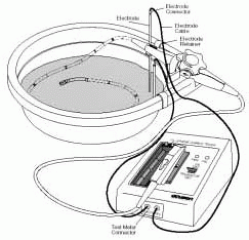
FIGURE 8.1. Proper technique for performing a leakage current test.


SAFETY

Serious complications associated with the use of TEE are rare (Table 8.3). The largest safety study reviewed data from 10,419 TEE procedures performed in awake patients (8). Morbidity occurred in 0.18% of exams: one death was reported. The safety of intraoperative TEE was
examined in a case series of 7200 cardiac surgical patients (9). No deaths related to intraoperative TEE were reported in this retrospective study. Morbidity occurred in 14 patients (0.2%); severe odynophagia accounted for half of these complications. In two other large series, morbidity was reported in 0.47% of 1500 ambulatory adult patients and 0% of 5016 cardiac surgical patients (10,11). The safety of TEE appears comparable with upper gastrointestinal (UGI) endoscopy (12).


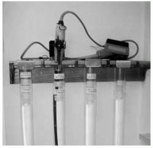
FIGURE 8.2. Example of a wall-mounted rack designed to protect transesophageal echocardiography probes during storage.


TABLE 8.3. Complications of Intraoperative TEE


Complications Related to the Gastrointestinal System


Esophageal perforation


Esophageal bleeding


Dysphagia


Odynophagia


Thermal injury


Transient bacteremia


Lip and dental trauma


Complications Related to Compression of Adjacent Structures


Tracheal compression


Displacement of the endotracheal tube


Vocal cord paralysis


Cardiac arrhythmias


Hypertension or hypotension


Splenic injury


Jul 15, 2016 | Posted by in CARDIOLOGY | Comments Off on Organization of an Intraoperative Echocardiographic Service: Personnel, Equipment, Maintenance, Safety, Infection, and Continuous Quality Improvement

Full access? Get Clinical Tree

Get Clinical Tree app for offline access