, Cesare Cuspidi2 and Sverre E. Kjeldsen3, 4
(1)
University of Milano Bicocca–IRCCS Istituto Auxologico, Italiano, Milan, Italy
(2)
University of Milano-Bicocca – Dept. of Health Sciences-IRCCS Istituto Auxologico Italiano Centro Ricerche Cliniche, Italiano, Milan, Italy
(3)
Department of Cardiology, University of Oslo, Faculty of Medicine, Ullevaal Hospital, Oslo, Norway
(4)
Division of Cardiovascular Medicine, University of Michigan Health System, Ann Arbor, Michigan, Norway
20.1 Introduction
A large number of studies in experimental animals and man show that angiotensin II, catecholamines and many other substances with cardiovascular and/or metabolic effects can favour alterations of organ function and structure independently on their ability to modify blood pressure (BP). There is, however, also a large body of evidence that these alterations are related to BP levels “per se” both in untreated and treated hypertensive patients. This chapter will offer some examples of this relationship mainly based on past work by the authors. The examples will largely focus on organ damages of more common assessment in clinical practice as well as of a more clear prognostic value, i.e. left ventricular hypertrophy, increased urinary protein excretion or reduction of glomerular filtration rate and carotid intima-media thickening or plaques. However, other types of organ damage will be also mentioned, albeit more briefly.
20.1.1 BP and Cardiac Damage
20.1.1.1 Left Ventricular Hypertrophy (LVH)
Both when assessed by echocardiography and when determined by electrocardiographic criteria, the prevalence of LVH increases progressively with the increase in office systolic or diastolic BP [1–4]. Evidence is also available that in hypertensive patients a BP reduction is associated with LVH regression regardless of the drug used to obtain BP-lowering effect [5], a greater BP fall being accompanied by a greater and more spread-out reduction in the markers of the abnormally elevated values of left ventricular volume, thickness and mass [6]. This is exemplified in Fig. 20.1, which refers to the large number of hypertensive patients recruited for the Losartan Intervention For Endpoint reduction (LIFE) trial. Both when the increase of left ventricular mass was assessed by Cornell voltage and when it was assessed by Sokolow-Lyon electrocardiographic criteria, the percentage of patients undergoing a LVH regression 12 months into the study became progressively greater as the treatment-reduced systolic or diastolic BP fall increased. Similar observations have been made for echocardiographic estimations of LVH regression and for treatments different from those (losartan or atenolol) used in the LIFE trial [7].
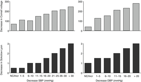
Fig. 20.1
Reduction in the incidence of left ventricular hypertension as a function of the treatment-induced reduction of systolic (S) and diastolic (D) blood pressure (BP) in the patients of the LIFE study. Left ventricular hypertrophy was assessed by electrocardiographic criteria, and the graphs show average values after 12 months of treatment (Kjeldsen)
20.1.1.2 Other Measures of Cardiac Damage
Although many more studies have focused on LVH, evidence is also available that other measures of cardiac damage exhibit a relationship with BP. One, observational studies have consistently shown that the diameter of the left atrium, the prevalence and the severity of systolic left ventricular dysfunction and the prevalence or the severity of diastolic dysfunction are both related to BP values [8, 9]. Furthermore, a treatment-induced BP reduction has been repeatedly shown to improve these measures of cardiac damage, irrespectively of the antihypertensive drugs used and independently on the concomitant modifications of other biomarkers with potential cardiac effects [10, 11]. A pertinent example is offered by the Study on Ambulatory Monitoring of Pressure and Lisinopril Evaluation (SAMPLE), in which hypertensive patients with echocardiographic LVH exhibited not only a LVH regression but also a distinct improvement of LV contractile performance, assessed by a sensitive marker such as fractional midwall shortening [12], after 1 year reduction of office and out-of-office BP reduction by treatment (Fig. 20.2) [13]. Interestingly, the improvement was entirely maintained after BP returned to the initially elevated values over a final month of placebo administration, indicating that the contractile function had not improved because of the favourable effect of a reduced afterload on cardiac performance but it had a persistent structural and/or functional basis (Fig. 20.2) [14].
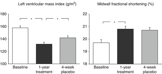
Fig. 20.2
Regression of left ventricular hypertrophy and improvement of left ventricular systolic function (midwall fractional shortening) with 24 h BP reduction over 1 year antihypertensive treatment in patients with hypertension and left ventricular hypertrophy. The left ventricular systolic function improvement was maintained when BP increased towards the initial baseline values after a final month of administration of placebo. From left to right histograms show baseline, 1 year treatment and final placebo data, respectively (From [14], with permission)
20.1.2 BP and Renal Damage
20.1.2.1 Microalbuminuria and Proteinuria
A huge number of studies have provided conclusive evidence that the prevalence of microalbuminuria or proteinuria is closely related to BP values, this being the case both in the general population and in diabetic or hypertensive nondiabetic patients [15, 16]. The evidence is also conclusive that a BP reduction by treatment is accompanied by a reduction of urinary protein excretion, regardless of the type of treatment employed, including that based on drug classes which also have a BP-independent antiproteinuric effect [17]. This is shown in Fig. 20.3 which refers to patients under treatment with drugs capable or not capable (irbesartan and amlodipine, respectively) to lower urinary protein excretion independently on BP changes. In both instances, the amount of urinary protein decreased progressively with the BP decrease, a similar phenomenon being apparent in the control group [18]. A further example is provided by the patients of the Ongoing Telmisartan Alone and in Combination with Ramipril Global Endpoint Trial (ONTARGET) in whom treatment consisted of an ACE inhibitor, an angiotensin receptor antagonist or a combination of the two drugs. In the patients pooled, the incidence of new-onset microalbuminuria or proteinuria was closely related to systolic BP down to very low on-treatment values, whose achievement was conversely associated with a more frequent return to normo-albuminuria in patients in whom an increased urinary protein excretion was detected at baseline (Fig. 20.4) [19].
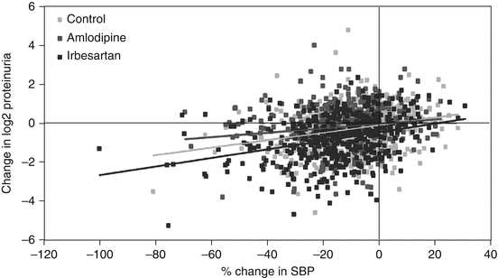
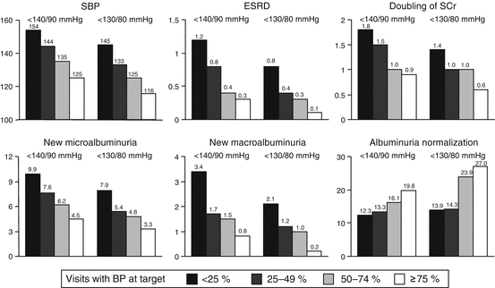
Fig. 20.3
Change in proteinuria associated with change of systolic BP after 1 year treatment in control patients as well as in patients treated with irbesartan or amlodipine. Regression lines between changes in proteinuria and BP are also shown (From [18], with permission)
Fig. 20.4
Incidence of renal endpoints according to the % of on-treatment visits before the renal event in which BP was reduced to < 140/90 mmHg or < 130/80 mmHg. The greater the % of visits with BP at target, the lower the mean SBP over the treatment period (upper right panel). SBP systolic blood pressure, ESRD end-stage renal disease, SCr serum creatinine (Modified from [19], with permission)
20.1.2.2 Reduction of Glomerular Filtration Rate
Observational studies have shown that in the general population, in diabetic patients and in patients with chronic kidney disease, glomerular filtration rate (GFR) as estimated (e) by conventional formulae [20] becomes progressively less as BP progressively increases [21]. It has further been shown that although the reduction of renal perfusion pressure may raise serum creatinine in the first few weeks after initiation of antihypertensive treatment, in the long term lower on-treatment BP values favour preservation of renal function and delay progression to end-stage renal disease in hypertensive patients with or without chronic nephropathy. This is again shown in Fig. 20.4 again for the normotensive and hypertensive patients recruited for the ONTARGET trial [19]. It has further been documented for the patients with hypertension and diabetic nephropathy addressed in a variety of studies in which the time-related loss of eGFR was plotted vs the achieved BP values [22]. As shown in Fig. 20.5, the lower was the on-treatment BP, the lesser was the decline of renal function as assessed by a reduction in GFR, the most effective renal protection being observed for mean arterial pressures (diastolic + one third of systolic) of about 100 mmHg, i.e. a value approximately corresponding to 120 mmHg systolic and 75 mmHg diastolic BP.
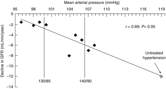
Fig. 20.5
Decline in glomerular filtration rate (GFR) according to on-treatment mean arterial pressure values in studies on diabetic nephropathy. Compared to untreated hypertension, antihypertensive treatment was associated with lower degrees of GFR decline. The smallest decline occurred at on-treatment mean arterial BP around 100 mg (From [22], with permission)
20.1.3 BP and Vascular Damage
20.1.3.1 Carotid Intima-Media Thickness
As exemplified in Fig. 20.6, thickening of the common or internal carotid artery walls increases progressively as BP increases [23]. Furthermore, evidence has been obtained that when BP is reduced by treatment, the progressive increase of carotid wall thickness that occurs with ageing and type 2 diabetes can be delayed and that some reduction may sometimes occur [24, 25]. Although the amount of evidence is not comparable with that available for LVH and renal damage, this justifies the conclusion that vascular structural alterations that reflect atherosclerosis are also in direct relationship with BP values. This is strengthened by the evidence obtained with the use of more direct measures of atherosclerotic lesions such as that obtainable by intravascular quantification of the volume of an atherosclerotic plaque (IVUS). The intravascular measured coronary plaque volume showed a continuous relationship with the BP changes induced by antihypertensive treatment supporting the notion of a direct antiatherogenic effect of BP-lowering interventions [26].
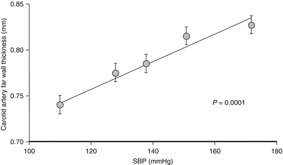
Fig. 20.6
Relationship between carotid artery far wall thickness and systolic blood pressure (SBP) in 1006 normotensive and hypertensive subjects (From [23] with permission)
20.2 Office or Out-of-Office BP?
Several studies have shown that in hypertensive patients cardiac, renal and vascular damages are more closely related to ambulatory than to office BP values. To quote some examples, both the prevalence of LVH and the echocardiographic left ventricular mass index value have been repeatedly found to have a higher correlation coefficient with the 24 h mean BP than with office BP values (Table 20.1) [27]. The prevalence of microalbuminuria and proteinuria has been reported to have a better association with 24 h mean than to office BP [28, 29], the correlation being closer for the night than for the daytime values [30]. Closer relationships with ambulatory than office BP values have finally been reported for carotid intima-media thickness [31, 32], the superior organ damage-ambulatory BP association extending in other studies to additional types of vascular-dependant organ damage, including those reflecting (1) small-artery functional or structural abnormalities [33] and (2) functional or structural brain alterations such as decline of cognitive function or white matter lesions [34, 35].
Table 20.1
Relationship of left ventricular mass index with office (or clinic) and 24 h mean systolic blood pressure (SBP)
Study | N | Clinic SBP | 24 h SBP |
|---|---|---|---|
Rowlands | 50 | 0.45 | 0.60 |
Drayer | 12 | 0.55 | 0.81 |
Devereux | 100 | 0.24 | 0.38 |
Lattuada | 50 | 0.07 | 0.40 |
Palatini | 42 | 0.52 | 0.62 |
Gosse | 61 | 0.40 | 0.49 |
White | 30 | 0.13 | 0.54 |
Gosse | 23 | 0.60 | 0.72a |
Prisant | 55 | 0.33 | 0.50 |
Verdecchia | 150 | 0.44 | 0.57 |
Ravoglib | 45 | 0.38 | 0.47 |
Its closer association with organ damage has strongly contributed to the current notion that ambulatory BP is prognostically superior to the BP derived from measurement in the physician’s office, presumably because ambulatory values reflect more accurately the BP regimen existing in daily life. Limited information exists, however, on the ability of out-of-office BP to predict progression or regression of organ damage over long-term longitudinal studies, although the available data also speak in favour of a greater ability of out-of-office BP, or of the treatment-induced out-of-office BP changes, to predict patients’ cardiovascular risk. In the hypertensive patients of the SAMPLE study, for example, the regression of LVH that occurred over 1 year antihypertensive treatment was more closely related to the reduction of 24 h mean than to office BP, the home BP reduction lying approximately in between (Fig. 20.7) [13]. Similarly, in the hypertensive patients of the European Lacidipine Study on Atherosclerosis (ELSA) and the Plaque Hypertension Lipid-Lowering Italian Study (PHYLLIS), the changes in left ventricular mass and carotid intima-media thickness, respectively, were significantly predicted by changes in ambulatory but not by changes in office BP [36, 37]. Finally, in the type I diabetic patients studied by the Valencia group, not only was the presence of proteinuria associated with a higher ambulatory BP value (Fig. 20.8), but a higher ambulatory BP value predicted the progression to microalbuminuria and nephropathy in individuals who did not have renal abnormalities at the start, independently on the office BP values [38].
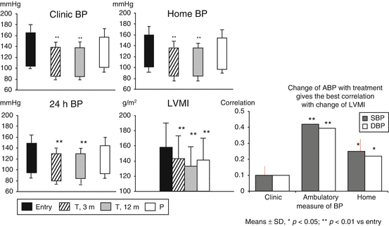
Fig. 20.7


Reduction from entry values of clinic (or office) self-measured home and 24 h (h) mean systolic blood pressure as well as of echocardiographic left ventricular mass index (LVMI) after 3 and 12 months of antihypertensive treatment in hypertensive patients with left ventricular hypertrophy. Data are also shown after a final month of placebo (P). Correlations between different BP and LVMI changes are shown at right BP blood pressure; A ambulatory (From [13] with permission)
Stay updated, free articles. Join our Telegram channel
Full access? Get Clinical Tree
