Open and Closed Mitral Commissurotomy for Rheumatic Mitral Stenosis
Bhagawan Koirala
Introduction
Rheumatic heart disease is still very common in the developing world. It affects mostly children and young adults. The pathophysiology of rheumatic fever and rheumatic heart disease is well described in the literature. The antibodies formed against the streptococcal antigen also attack the endocardial tissue (molecular mimicry), thereby causing inflammation of the target tissue. It involves the endocardium and the valves of the heart, joint surfaces and serosal surfaces of the pericardium, and pleura. Most commonly involved valves in order of frequency are mitral, mitral and aortic valve combined, and aortic valve alone followed by tricuspid valve. The pulmonary valve is almost never involved by the rheumatic process, and so, any involvement of this valve is considered to be of congenital origin unless proven otherwise.
The initial stage of mitral valve involvement always starts from at least some degree of mitral regurgitation (MR). That is because of chordal lengthening and some degree of associated myocarditis. As the process progresses the leaflets start to adhere to each other, thereby creating stenosis. In general, mixed stenotic and regurgitant lesions are more common than pure mitral stenosis (MS).
The symptoms and signs of mitral stenosis are similar to those of any mitral valve disease. Palpitations, exertional dyspnea, and occasionally, hemoptysis are the commonest symptoms at presentation. Paroxysmal nocturnal dyspnea (PND) and orthopnea may be present in severe cases. Typical examination findings are soft tapping apex beat, presence of an opening snap, and middiastolic rumbling murmur with presystolic accentuation. The presence of an opening snap usually denotes that the valves are pliable and indicative of a likely successful commissurotomy: open or closed.
As the stenosis becomes tighter the murmur generally increases in intensity and duration. However, with increasing fibrosis of the valve apparatus, the audibility of the opening snap and intensity of the murmur may decrease. Chest x-rays are a routine tool of investigation and give important information. In isolated mitral stenosis the left heart border is straightened because of left atrial appendage enlargement, but the left ventricle
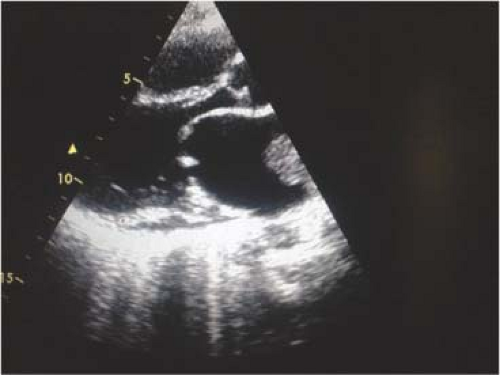
looks normal in size. Left atrial enlargement also leads to a double contour shadow in the right heart border and splaying of the carinal angle. The rhythm can be sinus or atrial fibrillation in many cases. Echocardiography is the mainstay of diagnosis and can give full information of the anatomy and function of the mitral valve. A transthoracic echocardiography can demonstrate the valve orifice area, degree of valve thickening, involvement of the subvalvar apparatus, the presence of mitral regurgitation, and left atrial thrombus, if any (Fig. 19.1). If there is any doubt about the presence of a left atrial thrombus, a transesophageal echocardiography (TEE) may be required.
looks normal in size. Left atrial enlargement also leads to a double contour shadow in the right heart border and splaying of the carinal angle. The rhythm can be sinus or atrial fibrillation in many cases. Echocardiography is the mainstay of diagnosis and can give full information of the anatomy and function of the mitral valve. A transthoracic echocardiography can demonstrate the valve orifice area, degree of valve thickening, involvement of the subvalvar apparatus, the presence of mitral regurgitation, and left atrial thrombus, if any (Fig. 19.1). If there is any doubt about the presence of a left atrial thrombus, a transesophageal echocardiography (TEE) may be required.
The American Heart Association/American College of Cardiology have defined four stages of rheumatic mitral stenosis:
Stage A: At risk of mitral stenosis: mitral valve doming during diastole, but normal transmitral flow velocity.
Stage B: Progressive mitral stenosis: progressive commissural fusion, doming of the valve with a mitral valve area (MVA) more than 1.5 cm2.
Stage C: Asymptomatic severe (or very severe) mitral stenosis: commissural fusion, diastolic doming with MVA <1.5 cm2 (or <1 cm2).
Stage D: Symptomatic severe (or very severe) mitral stenosis: commissural fusion, diastolic doming with MVA <1.5 cm2 (or <1 cm2), and symptoms of heart failure.
The above mentioned parameters constitute the main basis for indication of intervention. However, other factors such as the quality of valve leaflets, involvement of the subvalvar apparatus, and presence of thrombus are important considerations in selection of a surgical procedure.
Closed Mitral Commissurotomy
Closed mitral commissurotomy (CMC) is one of the early surgical procedures performed in the history of cardiac surgery and has given quality life to many of the patients.
CMC used to be a very common procedure in many countries in the past but now has been almost entirely replaced by percutaneous transcatheter balloon dilatation (PTBD).
The typical indications for CMC or PTBD are:
Symptomatic severe mitral stenosis with MVA less than 1.5 cm2
Asymptomatic very severe mitral stenosis with MVA of less than 1 cm2
Asymptomatic severe mitral stenosis with MVA <1.5 cm2 with new onset of atrial fibrillation
Mitral stenosis with history of PND or pulmonary edema.
Echocardiographic mean diastolic gradient across mitral valve more than 10 mm Hg is indicative of significant mitral stenosis but not considered as an indication independently because of its variability.
The only indication for CMC in the current era could be the need for commissurotomy when there are no PTBD facilities. It may also be performed when balloon dilatation is not feasible in small children. Mitral stenosis with failed PTBD is also an indication for CMC but usually requires an open procedure.
Detailed preoperative study of valve anatomy with 2D and color flow echocardiography are mandatory. Echocardiography will demonstrate the quality of the valve tissue, degree of stenosis, presence of subvalvar involvement, presence of left atrial thrombus, size of the left atrium, left ventricular function, and degree of mitral regurgitation. Wilkins score is a useful guide to identify patients suitable for a closed or open procedure. This is calculated based on the severity of changes in each component of the mitral valve apparatus, namely leaflet thickening, calcification, leaflet mobility, and involvement of the subvalvar apparatus. Wilkins score of less than 8 out of total 16 offers high chance of having a successful closed procedure.
The contraindications for a closed procedure (CMC or PTBD) are:
Presence of left atrial thrombus
Presence of more than mild mitral regurgitation
Presence of severe subvalvar involvement
Heavy calcification of the valve
Slight calcification of the tips of the leaflets is not a contraindication. Presence of a giant left atrium is a relative contraindication for CMC. Presence of atrial fibrillation per se is not a contraindication but needs detailed and careful evaluation for the possible presence of a left atrial thrombus.
Routine blood work including complete blood count, renal function test, serum electrolytes, routine urine examination, ECG, and chest x-ray are done before procedure. Echocardiography is performed a day before surgery to ensure that no new thrombus has formed in the left atrium. If a patient is on warfarin, it needs to be stopped for about a week before the procedure and have the international normalized ratio (INR) below 1.5. Medicines including beta-blockers or digoxin are continued until the day of surgery. While obtaining informed consent, the patient and the family need to understand that this procedure is not a curative one, and restenosis or regurgitation in the future are possible.
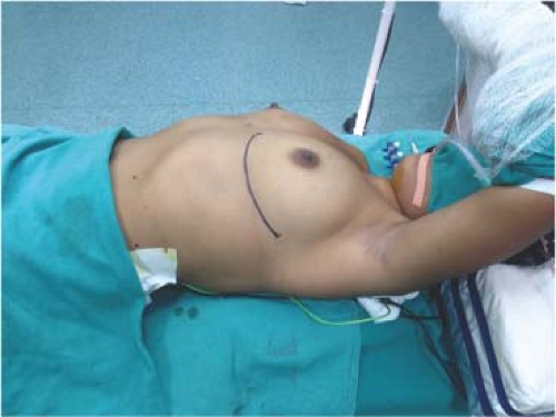
The patient is placed in a semilateral position with the left shoulder lifted approximately at 45 degrees and left hip slightly lifted with a sand bag. The left arm is lifted off and supported to a screen bar in a relaxed position (Fig. 19.2). A routine skin preparation and draping is done.
An anterolateral thoracotomy is performed along the fourth or fifth intercostal space. In females the breast tissue is carefully mobilized off the chest wall. The left lung is retracted and a vertical pericardiotomy is made about an inch anterior to the left phrenic nerve. The incision should reach the diaphragm below and main pulmonary artery above. Three to four stay stitches are placed along each of the margins of
the incised pericardium. The left atrial appendage and left ventricle come into perfect view at this time.
the incised pericardium. The left atrial appendage and left ventricle come into perfect view at this time.
Stay updated, free articles. Join our Telegram channel

Full access? Get Clinical Tree


