Fig. 29.1
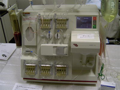
Under general anesthesia, patient is in prone position and bone marrow cells are collected from the iliac crest for about 1 h
The harvested bone marrow cells are diluted with RPMI 1640 (Gibco, Invitrogen, Carlsbad, CA, USA) containing heparin, and about 750 mL is saved in a sterile pack from the bone marrow collection kit (Baxter, Chicago, IL, USA). The mononuclear cell fraction is sorted and concentrated to a final volume of 100 mL with a COBE Spectra Apheresis System (Gambro, Stockholm, Sweden). The total count of mononuclear cells is 3.5–6.5 × 10⁹ cells. Next, we use the Isolex 300i Magnetic Cell Selection System (Nexell Therapeutics, Irvine, CA, USA) to separate CD34+ cells from the fraction of mononuclear bone marrow cells (Fig. 29.2). The final count of isolated CD34+ cells is 2.0–3.0 × 10⁷ cells.


Fig. 29.2
Isolex300i magnetic cell selection system separates CD34+ cells from the fraction of mononuclear bone marrow cells for about 3 h
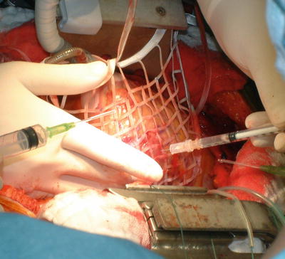
Coronary artery bypass is performed in our standard off-pump fashion. When the OPCAB procedure is completed, implantation of CD34+ cells into the infarction site is performed while the beating heart is stabilized with a heart net (Vital, Tokyo, Japan) (Fig. 29.3). This heart net can provide precise and equal injection of cells on the beating heart. The injection placement is based on preoperative echocardiography and SPECT viewing.


Fig. 29.3
A 5-mL cell suspension containing 2.0–3.0 × 10⁷ CD34+ cells is injected into 25–30 sites (0.2 mL to each site) with a 1 × 1-cm grid and a 26-gauge needle apparatus (6 mm deep)
A 5-mL cell suspension containing 2.0–3.0 × 10⁷ CD34+ cells is injected into 25–30 sites (0.2 mL to each site) with a 1 × 1-cm grid and a 26-gauge needle apparatus (6 mm deep). Using small needle can prevent bleeding at the puncture sites. After implantation, ventricular pacing wires and drainage tubes are put in place, and the chest wall is closed in standard fashion.
In our clinical study, we used CD34+ cells separated from bone marrow cells using an automatic mononuclear separating system. Other clinical studies of this therapy have used original bone marrow stem cell, bone marrow mononuclear cell, and CD133+ cell. Techniques of bone marrow collection are almost the same by all studies. Although methods of cell transplantation varied in each study, a direct intramyocardial injection of bone marrow cells is a most popular technique in the combination therapy of bone marrow cell transplantation and CABG. Some studies reported a method to inject bone marrow cells from a bypass graft; however, from the view point of cell delivery into insufficient vascular area, intramyocardial stem cell direct injection during surgery seems better. Furthermore, this therapy can accomplish all procedures on beating heart, at one operation room and in a day even if we take either methods of cell transplantation.
29.2.3 Follow-Up
After operation, patients are transferred to the intensive care unit for monitoring. Especially, it is very important to pay careful attention to new developing polymorphic premature ventricular contraction or ventricular tachycardia caused by cell transplantation. Before discharge, routine blood examination including cardiac enzymes and chest roentgenogram is taken. To evaluate short-term safety and feasibility of this therapy, electrocardiogram (ECG), transthoracic echocardiography, SPECT study, and cardiac catheterization including coronary artery assessment are routinely performed on the 1 month postoperative day. In our experiences, echocardiography and SPECT study are assessed on the 6, 12, 24, and 36 months compared with baseline values to evaluate mid- or long-term effectiveness of this therapy. The values (infarction wall thickness, LV end-systolic diameter, LV end-diastolic diameter) measured by M-mode echocardiograph are mostly available to assess the improvement of LV function.
Many papers which investigated the clinical effectiveness of this therapy also used echocardiography and SPECT study as evaluation methods of midterm outcomes [3–8]. Regarding SPECT study, we used stress ATP SPECT-sestamibi perfusion scans to determine myocardial perfusion during follow-up compared with baseline control. A few investigations used thallous chloride Tl-201 SPECT to assess the improvement in myocardial perfusion viability after intervention. Even if either radio nuclides are used, SPECT study is available methods to evaluate myocardial blood perfusion, distribution, and viability.
In current studies of this therapy, utilization of cardiac magnetic resonance imaging (MRI) and fluorine 18-fluorodeoxyglucose (18F-FDG) positron emission tomography (PET) has been reported. These methods may become useful in the future to evaluate a true effectiveness of postoperative cell transplantation except the influence of CABG [8, 9].
29.2.4 Outcomes
For the past 10 years, a large number of clinical studies in this bone marrow cell transplantation therapy have demonstrated their clinical benefits. Some RCTs compared with the combination therapy of cell transplantation and CABG with CABG alone also have showed their safety and effectiveness with excellent outcomes [3–9].
29.2.4.1 Intraoperative and Early Postoperative Outcomes
Regarding cell collection methods, there are some differences in each manipulation, but the number of harvested bone marrow stem cells is almost the same. In a few studies, there were the cases that required an intra-aortic balloon pump or a left ventricular assist device postoperatively, because these studies treated severe cases with low cardiac function. Few complications including fatal arrhythmia due to injection of cell transplantation did not present.
29.2.4.2 Echocardiography
Most studies of this therapy have demonstrated the significant improvement of LV function with echocardiography compared with their baseline data [3–9]. In RCTs, the improvement of cardiac function by echocardiography has been described as an increase in left ventricle ejection fraction (LVEF) of approximately 10 % in patients of combined therapy with CABG (Table 29.1). A few studies of combining cell transplantation with OPCAB reported similar results which implicates that cardiac arrest is not necessarily to perform safe and efficient cell implantation [3]. Moreover, this therapy showed a trend toward reduction in left ventricular end-diastolic volume (LVEDV) compared with control group. Yerebakan and colleagues reported that patients with LVEF not greater than 40 % had a possibility of improvement in LV function than patients with LVEF greater than 40 % in a mean follow-up of 65 months [9]. Our small study also presents an increase in LVEF of approximately 10 % in patients with cell transplantation within 1 month. It is very interesting knowledge from most clinical studies that the cardiac function in cell therapy patients significantly improves within 1 month in comparison with the baseline data [3–8].
Table 29.1
Treatment results of randomized controlled studies in bone marrow cell transplantation combined with coronary artery bypass grafting
Study author (ref) (year) | Sample size | Study design | Treatment group | Control group | Follow-up (mo) | LVRF/LVEDV evaluation | Change of LVEF (%) | Change of LVEDV (ml) | ||
|---|---|---|---|---|---|---|---|---|---|---|
Treatment | Control | Treatment | Control | |||||||
Patel et al. [3] (2005) | 20 | RCT | CD34 BMSC 22 × 106 | CABG onlya | 6 | Echocardiography | 16.7 ± 3.2 | 6.5 ± 1.8 | −22 ± 27.6 | −5 ± 12.4 |
Hendrikx et al. [4] (2006) | 20 | RCT | BMC-MN 60.25 ± 31.35 × 10⁶ | CABG only | 4 | Cardiac MRI | 6.1 ± 8.6 | 3.6 ± 9.1 | – | – |
Stamm et al. [5] (2007) | 40 | RCT | CD133 BMSC 5.8 × 10⁶ | CABG only | 6 | Echocardiography | 9.7 ± 8.8 | 3.4 ± 5.5 | −11.1 ± 38.6 | −4.4 ± 19.2 |
Ang et al. [6] (2008) (IC) | 63 | RCT | BMSC 84 ± 56 × 106 | CABG only | 6 | Cardiac MRI | −0.5 ± 1.8 | 0.7 ± 1.9 | 9.9 ± 14.64 | 17.9 ± 15.2 |
Ang et al. [6] (2008) (IM) | 63 | RCT | BMSC 115 ± 73 × 106 | CABG only | 6 | Cardiac MRI | 4.3 ± 1.58 | 0.7 ± 1.9 | −19.3 ± 14.82 | 17.9 ± 15.2 |
Zhao et al. [7] (2008) | 36 | RCT | BMC-MN 22.0 × 10⁸ | CABG only | 6 | Echocardiography | 13.3 ± 9.2 | 3.9 ± 4.9 | – | – |
Hu et al. [8] (2011)
Stay updated, free articles. Join our Telegram channel
Full access? Get Clinical Tree
 Get Clinical Tree app for offline access

| ||||||||||