Obesity and Dyspnea on Exertion
A 65-year-old obese woman gets admitted to the hospital with complaints of shortness of breath and dyspnea on exertion.
A. An enlarged left ventricle and a right-to-left shunt
B. An enlarged right ventricle and a left-to-right shunt
C. An enlarged right atrium and right ventricle and a right-to-left shunt
View Answer
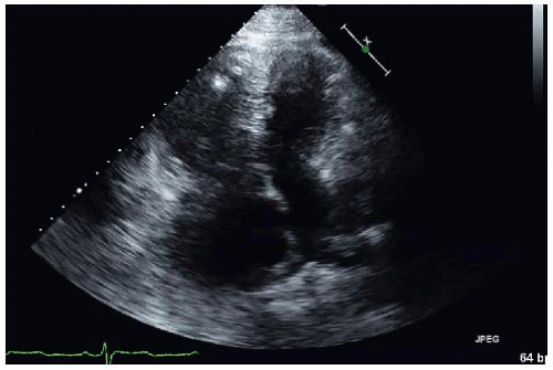
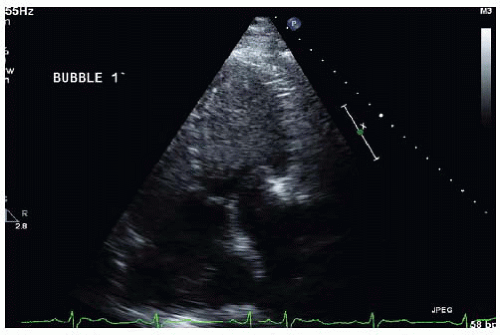
ANSWER 1: C. The transthoracic images are of fair quality due to the patient’s body habits. The first image shows a markedly enlarged right heart with right ventricular hypertrophy and the interatrial septum bulging to the right, consistent with both the patient’s pulmonary hypertension and elevated right atrial pressure. The second image shows opacification of the left ventricle with saline contrast, confirming a large right-to-left shunt. The opacification was almost immediate, but the exact site of the shunt was not visualized on this study.
QUESTION 2. The next best step would be:
A. A right and left heart catheterization
B. A transesophageal echocardiogram (TEE)
C. Placement of transcatheter atrial septal occluder device
D. A right heart catheterization with measurement of saturations
E. Start medical treatment of pulmonary hypertension
View Answer


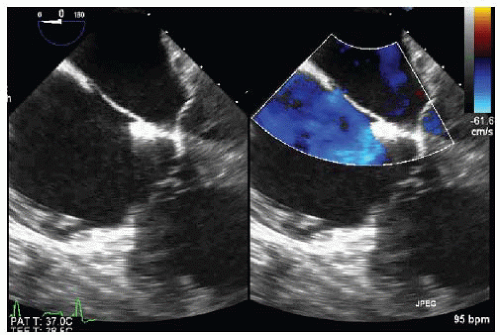
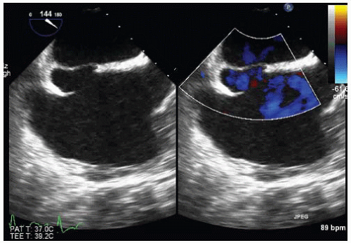
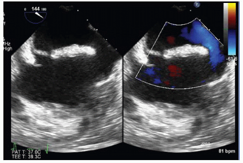
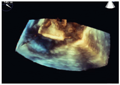
ANSWER 2: B. A TEE to identify the anatomy and site of the shunt would be a very reasonable first step. If a secundum atrial defect, which is the most common ASD, is detected then the TEE would help assess if the patient would be a candidate for a septal occluder device. There is no indication for a left heart catheterization. A right heart catheterization with saturations may still be indicated to evaluate the physiology of the shunt and evaluate pulmonary vascular resistance but would not clarify if the patient would be a candidate for percutaneous closure of an ASD. Correction of the intracardiac defect may be indicated rather than medical treatment of the pulmonary hypertension.
Stay updated, free articles. Join our Telegram channel

Full access? Get Clinical Tree


