Nuclear Cardiology

Chapter 7


Nuclear Cardiology


1. What is nuclear cardiology?


    Nuclear cardiology is a field of cardiology that encompasses cardiac radionuclide imaging, which uses radioisotopes to assess myocardial perfusion or myocardial function in different clinical settings, as well as radionuclide angiography, metabolic and receptor imaging, and positron emission tomography.


2. What is myocardial perfusion imaging (MPI)?


    MPI is a noninvasive method that uses radioisotopes to assess regional myocardial blood flow, function, and viability. The basis for MPI rests on the ability of this technique to demonstrate inhomogeneity of blood flow during stress compared with rest, thereby identifying ischemic or infarcted regions.


    During the stress portion of the test, exercise or a pharmacologic agent is used to produce vasodilation in the coronary vascular bed. Whereas a normal vessel can vasodilate and increase coronary blood flow up to four times basal blood flow, a significantly diseased or stenosed vessel cannot increase blood flow. Because radiotracer uptake into the myocardium is dependent on blood flow, the areas that are supplied by normal vessels in which there is maximally increased blood flow will take up more radiotracer than areas supplied by the stenosed vessel with relatively less blood flow. Therefore, there will be heterogeneous radiotracer uptake, which will be seen as a perfusion defect.


3. Define a perfusion defect and differentiate between a reversible and fixed defect.


    A perfusion defect is an area of reduced radiotracer uptake in the myocardium.


    If the perfusion defect occurs during stress and improves or normalizes during rest, it is termed reversible (Fig. 7-1). Generally, a reversible perfusion defect suggests the presence of ischemia.
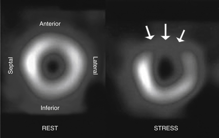


    If the perfusion defect occurs during both stress and rest, it is termed a fixed defect. Generally, a fixed defect suggests the presence of scar. However, in certain settings a fixed defect may not be scar. Instead, a fixed defect may represent viable tissue that is hibernating due to chronic significant or severe stenosis. Hibernating myocardium alters its metabolism in order to conserve energy. Therefore, it appears underperfused and has hypokinetic or akinetic function.


4. What are the different uses of MPI?


image MPI is used to diagnose coronary artery disease (CAD) in patients with intermediate risk for CAD who present with chest pain or its equivalent.


image MPI can be used to localize and quantify perfusion abnormalities or physiologic ischemia in patients with known CAD.


image MPI can be used to assess the presence of viability in areas of fixed defects using rest and redistribution studies with thallium.


image MPI can also be used for risk assessment and determination of prognosis with regard to cardiovascular events. It can be used as a prognostic tool in post–myocardial infarction patients, including patients with and without ST elevation, to identify further areas of myocardium at risk.


image MPI can also be used in the preoperative assessment to identify perioperative or postoperative cardiovascular risk. The extent and severity of perfusion defects are proportional to the risk of perioperative cardiac events.


5. Is MPI the most sensitive and specific test for diagnosing CAD?


    The ischemic cascade suggests that MPI would be a more sensitive test to detect ischemia because in the setting of ischemia, a perfusion abnormality occurs before a wall motion abnormality. The sensitivity of MPI is slightly better than stress echocardiography (85% versus 75%, respectively) and the specificity slightly worse (79% versus 88%). This results in similar accuracy for both types of stress tests. Both modalities are more sensitive and specific than treadmill or exercise electrocardiography testing, although according to the American Heart Association/American College of Cardiology (AHA/ACC) guidelines, the latter should be the first-line test for the diagnosis of CAD in someone who can exercise and has a relatively normal electrocardiogram (ECG).


6. List the different perfusion agents used in MPI.


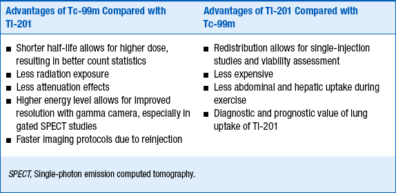
    For an agent to be an effective radiopharmaceutical, its distribution has to be proportional to regional blood flow, have a high level of extraction by the organ of interest, and have rapid clearance from the blood. The two most important physiologic factors that affect myocardial uptake of a radiotracer are variations in regional blood flow and the myocardial extraction of the radiotracer. In other words, there will be more uptake of a radiotracer in areas of increased blood flow and less in areas supplied by diseased or stenosed vessels. Importantly, because myocardial extraction is an active process with regard to thallium-201 and a mitochondrial-dependent process with regard to the technetium-99m agents, it can only occur if the cells in that region are viable. The relative advantages and disadvantages of thallium-201 and technitium-99 are summarized in Table 7-1.


Jun 5, 2016 | Posted by in CARDIOLOGY | Comments Off on Nuclear Cardiology

Full access? Get Clinical Tree

Get Clinical Tree app for offline access