Fig. 2.1
Right anterior oblique external view of the human heart (Photographs the courtesy of Dr William Edwards)
2.1.1 Venous Return to the Heart
The heart receives venous return both from the body and lungs, and from the heart itself. Systemic venous return from the body is through the superior vena cava and inferior vena cavae. Venous return from the lungs is via pulmonary veins, and venous return from the heart is via the coronary sinus as well as sinusoidal veins in the right ventricle.
Normally, the superior vena cava is a right-sided structure as is the inferior vena cava. Usually, there are four pulmonary veins, two from the left and two from the right; however, it is not abnormal to have a total of three or five pulmonary veins. The superior and inferior vena cavae empty into the right atrium. The pulmonary veins empty into the left atrium. The venous return from the heart muscle itself enters the right atrium through a coronary sinus, the os of which is in the right atrium and to the right ventricle via the coronary veins and right ventricular sinusoids.
2.1.2 Atria
There is a right atrium and a left atrium. These structures are not named just for their relative position in the chest but rather for their morphological features. A morphological right atrium normally is on the right side of the body, and a morphological left atrium normally is on the left side of the body. However, there are a number of cardiac anomalies in which the morphologically right atrium may be on the left side of the body and the morphologically left atrium may be to the right side of the body. A right atrium is characterized by the limbus of the fossil ovalis, a large pyramidal-shaped atrial appendage, the presence of the crista terminalis and pectate muscles, and the fact that it receives the vena cava and the coronary sinus. Alternatively, a left atrium is characterized by the presence of a small fingerlike atrial appendage, the absence of a crista terminalis, and the absence of pectinate muscles.
2.1.3 Atrioventricular Valves
There are two atrioventricular valves. The tricuspid valve allows blood to travel from the right atrium to the right ventricle, and the mitral valve allows blood to move from the left atrium to the left ventricle. The tricuspid valve, as its name implies, is a three-leaflet structure. The mitral valve, as its name implies, is a bileaflet structure. The annuli of both of these valves attach to the ventricular septum, but the mitral valve attaches in a more cephalad position than the tricuspid valve (Figs. 2.2 and 2.3). Hence, there is a septum which on one side is in the left ventricle and on the other side is in the right atrium. This is termed the “atrioventricular septum.” Another feature of a tricuspid valve is that it has a papillary muscle attached to the ventricular septum, but the mitral valve does not. Mitral valves do not have papillary muscles on the ventricular septum.
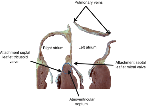
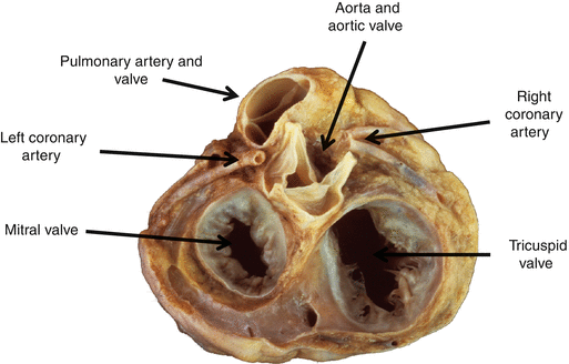
Fig. 2.2
Four-chamber view of a human heart. Note that the septal leaflet of the mitral valve attaches more cephalad than the septal leaflet of the tricuspid valve (Photographs the courtesy of Dr William Edwards)
Fig. 2.3
Pathologic specimen demonstrating the relationships of the four valves of the heart to each other and the location of the proximal coronary arteries (Photographs the courtesy of Dr William Edwards)
2.1.4 Ventricles
There is a right ventricle and a left ventricle. These ventricles are not named simply because the right ventricle is on the right and the left ventricle is on the left. In fact the right ventricle is anterior and to the right of the left ventricle. They are morphologically dissimilar. A right ventricle is a thinner-walled structure than the left ventricle. A right ventricle has an inflow portion, body, and an outflow portion. The distribution of muscle in the right ventricle is arranged such that the right ventricle is a more compliant chamber than the left ventricle. It is designed to accommodate changes in volume. The left ventricle is a thicker-walled left compliant chamber than the right ventricle. The orientation of its muscle fibers is such that it is designed to pump higher pressure than the right ventricle is designed to pump.
2.1.5 Semilunar Valves
There are two semilunar valves: the pulmonary valve and the aortic valve (Fig. 2.3). Both of these valves have three cusps. The pulmonary valve has muscle beneath it (a “conus”) such that the annulus of the pulmonary valve and the annulus of the tricuspid valve are not contiguous (Fig. 2.3). On the other hand, the aortic valve annulus is contiguous with the annulus of the mitral valve. This is an important distinction when dealing with a number of congenital anomalies of the heart. The aortic valve is oriented to the right and posterior to the pulmonary valve. The coronary arteries arise from the aorta in the region of the cusps of the aortic valve. There is dilation in the area of the cusps of the aortic valve, and these areas of dilation refer to sinuses of Valsalva. Similarly, there are dilations by the cusps of the pulmonary valve. These are called the sinuses of the pulmonary valve and should not be confused with the term sinuses of Valsalva.
2.1.6 Great Arteries
There are two great arteries, the pulmonary artery and the aorta. The pulmonary artery arises from the right ventricle and divides into the right and left pulmonary arteries. The aorta arises from the left ventricle. The initial branches of the aorta are the right and left coronary arteries. The next branch is the innominate artery that gives rise to the right carotid artery and right subclavian artery. The next branch of the aortic arch is the left carotid artery and the next branch is the left subclavian artery.
Stay updated, free articles. Join our Telegram channel

Full access? Get Clinical Tree


