Lower prevalence of coronary artery disease
Higher prevalence of non-ischaemic chest pain (microvascular abnormalities, mitral valve prolaps)
Less predictive symptomatology
Limited exercise tolerance due to older age at initial diagnosis
Different response to exercise than men
Limited exercise capacity (mostly due to older age)
Lower peak exercise values
Lesser increase in left ventricular ejection fraction
Increase in cardiac output by enhancing end-diastolic volume
Inappropriate catecholamine release
Hormonal influences of oestrongens mimicking digitalis-like false positive ECG response
Anatomical differences affecting stress test results
Female breast attenuation artefacts
Smaller coronary artery size
Smaller left ventricular chamber size
Higher prevalence of single vessel disease
Based on the Women’s Ischemia Syndrome Evaluation (WISE) study, the assessment of CAD in women should not just focus on epicardial disease but also on abnormal coronary reactivity, microvascular dysfunction and distal microembolization. These factors not only contribute to higher rates of angina and ischemia in women, but also to their clinical outcome.
This chapter focuses on the noninvasive evaluation of CAD in women. Various classic and novel methods of establishing risk and diagnosing CAD are discussed along with specific benefits and pitfalls of each approach. A proposed approach is included at the end.
Noninvasive Diagnostic Testing
Noninvasive testing is used in the diagnosis and risk stratification of CAD to better identify patients who would benefit from medical therapy or revascularization procedures, as well as to exclude CAD. Although substantial research efforts have been made to improve the diagnosis and treatment strategies for women at risk, the detection of CAD in women can be problematic. A great challenge in diagnostic testing in women is selecting the appropriate test for the patient. Establishing pre-test probability is imperative to achieving optimal incremental value in post-test information. Thus selection of candidates for diagnostic testing is based on Bayesian theory, in which the post-test likelihood becomes a function of the patient’s pre-test risk [5]. In low-risk women, the change from pre-test to post-test risk estimation is minimal. However, non-invasive testing is most useful in the intermediate-risk population, as it may shift women to lower-risk cohorts with negative test results, or higher-risk cohorts with abnormal results.
Early detection of ischemic heart disease is especially important in symptomatic women. As mentioned before, women with CAD experience worse outcomes than men, irrespective of age. These gender based differences exist as women tend to have more advanced disease, due to lack of early recognition and management. Accurately chosen noninvasive diagnostic tests can be useful in identifying women at the earliest stage of presentation so that appropriate therapeutic strategies can be implemented early on to improve the outcomes [4, 6, 7].
Establishing Risk
In the asymptomatic woman the goal of risk assessment is to identify those patients at increased risk of developing CAD. This can be achieved by various methods including patient history, risk factors, risk prediction scores, and biomarkers.
Various risk scores have been developed which incorporate numerous risk factors and attempt to stratify patients into low, intermediate or high risk categories. The Framingham risk score (FRS) is a global risk score which utilizes traditional risk factors for CAD including age, smoking, blood pressure, diabetes, and cholesterol values [8]. Women with low, intermediate, and high FRS have expected annual rates of CAD death or myocardial infarction of less than 0.6 % (low risk), 0.6–2.0 % (intermediate risk), and greater than 2 % (high risk), respectively. However, the FRS often underestimates risk in women [9]. In the Multi-Ethnic Study of Atherosclerosis, when excluding women with diabetes and those older than 79 years, 90 % of women were classified as “low risk” based on the FRS [10]. The use of coronary artery calcium scoring led to significant re-classification in this cohort.
The Reynolds risk score is a recently derived and validated gender-specific tool for predicting incident cardiovascular events (myocardial infarction, ischemic stroke, coronary revascularization, and cardiovascular death) [11]. The score includes age, systolic blood pressure, high-sensitivity C-reactive protein, total cholesterol, high density lipoprotein cholesterol (HDL), hemoglobin A1c, smoking status, and family history of premature myocardial infarction. When compared to the FRS, use of the Reynolds score resulted in risk reclassification in >40 % of intermediate FRS women.
The novel risk marker used in the Reynolds risk score, high-sensitivity C-reactive protein (hsCRP), has been shown to improve detection of CAD in women. Women on average have greater mean CRP measures from early age as compared to men [12]. It appears that this difference in CRP accounts for greater frequency of inflammatory mediated autoimmune diseases in women as compared to men, suggesting a possible role of inflammation in gender difference of CAD. Studies have shown that the relative risk of future cardiac events increases proportionally with increasing levels of hsCRP, thus accelerating risk [13, 14]. A position paper from the European Society of Cardiology noted that CRP is one of the best candidates for screening; however, evidence is lacking to recommend its routine use [15]. They noted that a CRP >3 mg/L reclassified only 5 % of intermediate risk women. Thus CRP has the greatest utility when is combined with the Reynolds risk score.
Several other risk factors are worth mentioning given the substantial gender-related variability in the prevalence and outcome associated with them. Hypertriglyceridemia is a potent independent risk factor for ischemic heart disease in women as compared to men [16]. In a meta-analysis of 17 studies hypertriglyceridemia increased the risk for coronary heart disease by 32 % in men and 76 % in women [17]. In a report from the WISE study, women with metabolic syndrome have an increased prevalence of subclinical disease and are at a two-fold higher relative risk for cardiac events as compared to women with a normal metabolic status [18].
Hormonal changes throughout a woman’s lifetime also appear to play a key role in their risk for ischemic heart disease. Ovarian dysfunction, as seen in functional hypothalamic amenorrhea as well as polycystic ovarian syndrome, has been shown to be associated with premature atherosclerosis [19, 20]. A recent meta-analysis found that preeclampsia doubles the risk for subsequent ischemic heart disease [21]. Thus, obtaining a gynecological and obstetrical history can aid in further risk stratification.
An interesting new tool on the horizon is a gene expression score (GES) which was recently developed and validated as a reliable diagnostic approach in the assessment of non-diabetic patients, especially women, with suspected obstructive CAD [22]. The algorithm uses gender, age, and the expression level of 23 genes which then generates a score on a 1- to 40-point scale, where increasing scores indicate increasing likelihood of obstructive CAD. As compared to symptom or myocardial perfusion imaging diagnostic approaches, the GES performed similarly in women and men. The GES may be especially helpful in the assessment of obstructive CAD in non-diabetic women for whom the use of symptoms and functional testing has proven unreliable. In future decades further advances in genetically based risk stratification will allow for more tailored gender-based evaluation of CAD risk.
Evaluation of Atherosclerotic Burden
The noninvasive measurement of atherosclerotic burden is another great tool for evaluating women at risk of adverse ischemic events. An established tool used in clinical practice is the ankle brachial index. Studies have shown that in women, the prevalence of an ankle brachial index ≤0.90 increases with age (approximately <5 % for women <60 years of age, up to 10–35 % in women 60–80 years of age) and is more prevalent in Black and Hispanic women [23, 24]. The 10-year cardiovascular mortality for an ankle brachial index ≤0.90 even after adjusting for the Framingham Risk score was significantly elevated in women (HR 3.0, 95 % CI 2.0–4.4) and men (HR 2.9, 95 % CI 2.3–3.7) [25].
Novel imaging techniques have allowed further evaluation of subclinical CAD in women. The roles of carotid intima media thickness (IMT) as well as presence of focal plaques detected by ultrasonography have been extensively studied as subclinical atherosclerotic markers. In women a low risk carotid IMT was associated with a ~1 % 10 year coronary heart disease risk versus ~10 % for a high risk carotid IMT [26]. A higher carotid IMT predicted a relatively greater risk of coronary heart disease for women than men [27].
The measurement of coronary calcifications using coronary CT is another useful marker of atherosclerotic disease burden. Coronary artery calcium (CAC) measurements, with either electron beam tomography or multidetector CT, are calculated and translated into a mean Agatston score which further places patients in various risk categories. In one study of 220 women with a normal coronary arteriogram, none had detectable CAC thus yielding a negative predictive value of 100 % [28]. However, women with moderate (≥100) or high (≥400) CAC scores had greater prevalence of obstructive coronary disease in the same study. In a large cohort of over 10,000 asymptomatic patients (>4,000 women), the extent of CAC was an independent and incremental estimator of all-cause mortality [29]. For women, as compared to a CAC score of ≤10, the risk-adjusted relative risk ratios for all-cause mortality were elevated according to increasing CAC values: 2.5 for a CAC value of 11–100, 3.7 for CAC of 101–400, 6.3 for a CAC of 401–1,000, and 12.3 for a CAC of >1,000 (p < 0.0001). Importantly, in this large cohort, for a given CAC score mortality rates were 3–5 fold higher for women as compared to men – thus suggesting a need for gender-specific cutoff values. Calcified plaque burden parallels overall plaque burden as CAC is almost always present in cases of angiographically significant CAD, and thus demonstrates great sensitivity but low specificity since detection of calcification implies atherosclerosis, but it is not specific for luminal obstruction and does not reveal the hemodynamic significance of the stenosis [4]. However, the test has a high negative predictive value when no calcifications are identified.
Exercise Stress Testing
The exercise ECG is the oldest and most commonly used form of stress testing. Multiple studies have shown profound gender differences when using treadmill ECG testing. According to the ACC/AHA exercise testing guidelines, women should undergo exercise testing if they are at an intermediate pretest probability for CAD on the basis of symptoms and risk factors, have a normal ECG, and are capable of maximal exercise [30]. However, exercise ECG testing has diminished accuracy in women. In a meta-analysis of over 3,000 women undergoing exercise ECG studies, the sensitivity and specificity were 61 and 70 % respectively for detection of obstructive CAD [31]. In similar studies, the mean sensitivity and specificity in men were 72 and 77 %, respectively [30].
Several factors account for gender based differences in the accuracy of exercise ECG testing. Women tend to have lower CAD prevalence and a greater prevalence of single-vessel disease as compared to men, they tend to be older when they present, and women have decreased exercise tolerance – all factors contributing to a decreased ability of the test to induce ischemia and ST depression thus limiting accurate identification of women with CAD [7]. Women more often have resting ST-T wave changes and lower ECG voltage [32].
Several studies have shown that significant exertional ST segment depression in women did not differ between those surviving vs. those dying from cardiovascular death during a follow up period of 20 years [33, 34]. However, marked ST segment changes (≥2 mm ST depression, horizontal or downsloping) occurring at low workloads or persisting into recovery confirmed high risk status for women [32].
The use of the Duke treadmill score (DTS) has been shown to improve the accuracy of exercise ECG testing in women. The DTS is defined as exercise time – (5 × ST deviation) – (4 × chest pain [1 = nonlimiting, 2 = limiting]). In a study of over 900 symptomatic women undergoing exercise ECG testing followed by coronary angiography, significant coronary stenoses (≥75 %) were present in 19, 35, and 89 % of low-, moderate-, and high risk women based on their DTS risk categories [35]. Another study of >5,000 asymptomatic women evaluated the association of the DTS and mortality [36]. They noted that after adjusting for the FRS, the risk of death decreased by 9 % for each unit increase in the DTS. Those women with a DTS <5 (moderate or high risk) had hazard ratios for death and cardiac death that were 2.2 and 2.5 times greater, respectively, than did those who had a DTS ≥5 (low risk), after adjusting for the FRS.
Functional capacity is a major predictor of adverse coronary events and all-cause mortality in both men and women. Women often have lower functional capacity as compared to men and have a greater functional decline with age [37]. Sedentary women are often incapable of performing greater than five metabolic equivalents (which are required to proceed to stage II of the aggressive Bruce protocol) due to excessive dyspnea and premature fatigue, thus leading to inadequate diagnostic information. In the abovementioned study of over 900 asymptomatic women, the investigators noted that the predictive value of the DTS was entirely due to the exercise component of the score. For each one metabolic equivalent increase in exercise capacity, the mortality was decreased by 17 % after adjusting for the FRS [36].
To better approximate performance during exercise ECG testing, it is important to estimate a woman’s ability to perform activities of daily living as a guide to approximate peak metabolic equivalent levels. The Duke Activity Status Index (DASI) is a brief, self-administered, 12 item questionnaire which evaluates a subjects’ ability to perform a variety of common activities of daily living, and was found to be a valid measure of functional capacity [38]. In the WISE study, two thirds of the cardiac events in women occurred in those with an estimated capacity of less than 4.7 DASI metabolic equivalents [39]. Women achieving less than 4.7 metabolic equivalents had a 3.7 fold increased risk of death or non-fatal myocardial infraction when compared to women reporting higher functional capacity. In the same study, each one metabolic equivalent increase in the DASI score was independently associated with an 8 % decrease in the risk of major adverse cardiovascular events. The current exercise testing guidelines support referral to pharmacologic stress testing with imaging in patients with submaximal exercise capacity [30].
The heart rate response further aids in risk stratification of women. The inability to reach 85 % of maximum age-predicted heart rate (HR) in women has been associated with a higher likelihood of CAD as well as with a decreased survival [40, 41]. Heart rate recovery in the first few minutes also improves risk assessment. In a study of 720 women undergoing exercise ECG testing and subsequent coronary angiography, 30 % had an abnormal HR recovery (defined as a decrease of HR 12 bpm or less in the first minute of recovery), which was independently predictive of death after adjustment for multiple covariates (HR 1.5, 95 % CI 1.2–1.9) [42]. Data on blood pressure response to exercise have been inconclusive in women.
Hormonal factors also play a role in exercise testing. Endogenous estrogen may have a digoxin-like effect thus promoting higher false positive exercise ECG rates in premenopausal women [3]. Angina and ischemia have been shown to vary by the menstrual cycle as well [43]. In the luteal/menstrual phase, where estradiol levels are low, a greater prevalence of ischemia and reduced time to ischemia onset have been noted. In postmenopausal women, hormone replacement therapy may result in false negative test results due to its vasodilatory properties [3].
Up until recently, there has been a lack of randomized controlled trials to guide diagnostic decisions in women thus resulting in variability of utilization patterns of various testing strategies. A recent randomized trial (What Is the Optimal Method for Ischemia Evaluation in Women [WOMEN] trial) of 824 symptomatic middle-aged (median = 62 years) women with suspected CAD evaluated the incremental value of myocardial perfusion imaging in clinical decision making over standard exercise ECG testing [44]. At 2 years of follow-up there was no difference in major adverse events (CAD death or hospitalization for acute coronary syndrome or heart failure) between both groups. Thus in low risk women, capable of exercise, the use of exercise ECG as the initial diagnostic strategy yielded similar outcomes to myocardial perfusion imaging but with significant diagnostic cost saving.
The exercise ECG is a commonly used form of stress testing; however, women who have an abnormal resting ECG, a decreased exercise capacity, or diabetes mellitus may need to undergo cardiac imaging with either exercise or pharmacologic stress.
Stress Echocardiography
Stress echocardiography is a useful tool not only for evaluation of stress induced ischemia but also for systolic and diastolic dysfunction, and the extent of ischemia and infarction. Assessment of valvular heart disease, or pericardial abnormalities can also help reveal an alternate explanation for symptoms of dyspnea or chest pain. Exercise echocardiography can be performed via a treadmill or bicycle, while for patients who cannot exercise, dobutamine is the most commonly used pharmacologic stress agent. Stress echocardiography has been shown to have high accuracy in detecting CAD in women.
Based on a compilation of meta-analyses on the diagnostic accuracy of stress echocardiography in women, the mean sensitivity was 84 % and mean specificity was 76 % [3]. When comparing exercise echocardiography to an exercise score (encompassing exercise ECG interpretation and exercise capacity and hemodynamics) in women, exercise echocardiography had higher sensitivity and specificity [45]. In a meta-analysis specifically comparing exercise ECG, exercise echocardiography, and exercise thallium for the diagnosis of angiographically confirmed CAD in women, the investigators found that exercise echocardiography had the highest sensitivity and specificity of all three modalities [31]. Although stress echocardiography has somewhat lower sensitivity for detecting intermediate stenoses or single-vessel CAD, its high negative predictive value makes it particularly useful test to exclude ischemia in younger women [46]. Exercise echocardiography appears to have the best balance between accuracy and cost for the diagnosis of coronary artery disease in women as compared to exercise ECG [47].
Dobutamine stress echocardiography (DSE) has been found to have a mean sensitivity of 80 % and specificity of 84 % in a large meta-analysis, both higher than pharmacologic stress testing with adenosine or dipyridamole [48]. One study of 306 patients (96 women), found that the overall specificity and the regional accuracy of DSE was higher in women than in men [49].
Stress echocardiography has been shown to be useful for determining cardiovascular prognosis in women. One study showed that the results of exercise echocardiography have comparable implications in both men and women when evaluating outcomes of cardiac death and nonfatal myocardial infarction [50]. A large study examined the 5-year mortality in 4,234 women undergoing exercise or dobutamine stress echocardiography [51]. Risk-adjusted 5-year survival was 99.4, 97.6, and 95 % for exercising women with no, single, and multi-vessel ischemia. Significantly worse survival was noted for women undergoing dobutamine stress, where 5-year survival was 95, 89, and 86.6 % for those with no, single, and multi-vessel ischemia. A more recent study of over 8,000 patients (3,208 women) undergoing stress echocardiography (exercise, dobutamine, and dipyridamole) evaluated its prognostic value of suspected and known CAD [52]. In patients with known CAD, women had a higher event rate than men in the presence of ischemia. The annual event rate of death or nonfatal MI was 7.0 % in women and 5.8 % in men with CAD (p = 0.08). The annual event rate was 2.4 % in women and 3.6 % in men with suspected CAD (p < 0.0001).
Limitations to stress echocardiography include poor acoustic windows due to obesity or breast tissue as well as lung disease; however lack of radiation makes it an attractive option. In women, stress echocardiography with exercise or dobutamine is an effective and accurate noninvasive method of detecting ischemia and risk stratifying symptomatic women with intermediate to high pretest likelihood of CAD.
Myocardial Perfusion Imaging
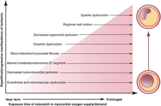
Changes in myocardial perfusion are one of the first manifestations in the time course of the ischemic cascade, preceding wall motion abnormalities, ECG changes, and symptoms (Fig. 4.1). Myocardial perfusion imaging may thus provide a more precise measure of estimating CAD risk. Myocardial perfusion can be evaluated in women using single-photon emission computer tomography (SPECT), positron-emission tomography, or cardiac magnetic resonance (CMR).
Nuclear Myocardial Perfusion Imaging
The most commonly nuclear based myocardial perfusion imaging technique used today is single-photon emission computer tomography (SPECT) which can provide information on perfusion defects, global and regional ventricular function, and left ventricular volumes.
The average diagnostic accuracy based on several meta-analyses of stress SPECT imaging in women reveals a sensitivity of 81 % and specificity of 66 % [3]. In a study evaluating patients with known and suspected CAD, when correcting for selection bias, the sensitivity and specificity results did not differ significantly between men and women [54]. Another study comparing the diagnostic accuracy of thallium-201 (Tl-201) with gated technetium-99m (Tc-99m) sestamibi SPECT in women noted that the sensitivity for detecting CAD was similar for both tests [55]. However, specificity improved dramatically from 67 % for Tl-201 to 92 % for gated Tc-99 sestamibi SPECT. When focusing on symptomatic postmenopausal women undergoing exercise or pharmacologic stress ECG gated SPECT imaging, the sensitivity and specificity were both 88 % [56]. In men and women with left bundle-branch block, vasodilator pharmacologic stress SPECT imaging has been shown to be more accurate than exercise perfusion imaging in the identification of CAD [57].
Multiple large studies have demonstrated the powerful predictive value of myocardial perfusion imaging in women. In a study of 3,402 women and 5,009 men with symptoms, stress SPECT imaging had a similar prognostic ability regardless of gender [58]. For women without ischemia the 3 year survival rates were 99 % as compared to 85 % for those with ischemia of three vessel territories. A normal myocardial perfusion study demonstrated an annual cardiac event rate of <1 % in pooled data from more than 7,500 women [5]. The abovementioned study of postmenopausal women undergoing SPECT imaging also evaluated 5-year occurrence of hospitalization for acute coronary syndrome, myocardial infarction and new onset or worsening angina [56]. Cox survival analysis showed a 5-year cumulative event-free survival rate of 94 % for patients with normal test results, as compared to 48 % for those with abnormal SPECT scans. Dual-isotope myocardial perfusion imaging yields incremental prognostic value in both men and women; however, it is able to identify high risk women more accurately than high risk men [59].
Diabetic women are a special high risk group of patients for which myocardial perfusion imaging is especially beneficial in predicting outcomes. In a study of men and women with and without diabetes, the survival rates (both for death and death or myocardial infarction) for diabetic women were the lowest for any amount of ischemia as compared to all other groups [60]. Cardiac event rates were significantly higher among the 451 diabetic women than in the 1,635 nondiabetic women (8 % vs. 3.2 %, respectively; p < 0.01). Another study of over 5,000 patients (~50 % women) showed that dual isotope adenosine SPECT imaging provided incremental value over pre-scan data for the prediction of cardiac death in both genders [61]. Similarly, this study illustrated that diabetic women and patients with insulin-dependent diabetes had a significantly greater risk of cardiac death than other patients for any SPECT result.
A recent study evaluated the effect of gender on the prognostic information obtained from left ventricular volume indices and ejection fraction in a SPECT studies [62]. ECG-gated SPECT studies were evaluated for end-systolic and diastolic volume indices as well as ejection fraction in 891 patients (43 % women), and were used to predict hard cardiac events as well as the combined endpoint of all-cause mortality or non-fatal myocardial infarction. The investigators noted that women had smaller left ventricular volume indices and higher ejection fraction despite equivalent rates of hard cardiac events and the combined endpoint mentioned above. Thus, in women, the risk of subsequent events starts at smaller volume indices compared to men despite similar risk profiles.
There are several important limitations to SPECT imaging in women [32]. Women have smaller hearts raising the possibility that smaller areas of reduced myocardial perfusion may be missed as a result of limitation in spatial resolution. Breast tissue attenuation and obesity can interfere with image quality and cause false positive results. As SPECT flow is comparatively assessed across the myocardium, it can appear normal in the setting of global reductions in perfusion such as multi-vessel CAD, diffuse endothelial or microvascular disease, left ventricular hypertrophy, or cardiomyopathy [53]. Lastly, radiation exposure is of concern. The addition of ECG gating, attenuation correction protocols, and use of higher energy radioisotope technetium (which allowed lower dosing) have made SPECT imaging less problematic in women. Newer techniques such as ultrafast cardiac SPECT cameras with cadmium-zinc-telluride (CZT) -based detectors are faster, produce higher quality images as compared to conventional SPECT cameras, and may allow lower mean radiation dose and shorter imaging time [63]. Recent reports of stress SPECT imaging with only 4–5 mCi Tc-99m dose and a 12–16 min acquisition time in non-obese patients have been feasible, thus decreasing the patient’s time in the imaging laboratory and decreasing the patient’s radiation exposure [64]. Use of stress only protocol with this camera in patients with symptoms, presence of cardiac risk factors, but no prior history of CAD, can allow ultra-low dose of radiation (only 1–2 mSv), which may be especially useful in women.
Stress myocardial perfusion imaging is recommended for symptomatic women with intermediate to high pretest likelihood of CAD. Intermediate risk women, who have normal test results are unlikely to have CAD and usually do not need further testing. A normal SPECT in women has an excellent negative predictive value of 99 % and carries a very low event rate (<1 %) [65, 66].
Table 4.2 presents a comparison of diagnostic accuracy of commonly ordered tests.
Table 4.2
Comparison of diagnostic accuracy of commonly ordered tests
Exercise electrocardiography | Stress echocardiography | Stress SPECT | ||||
|---|---|---|---|---|---|---|
Author, year (Ref.) | Sensitivity (%) | Specificity (%) | Sensitivity (%) | Specificity (%) | Sensitivity (%) | Specificity (%) |
Fleischmann et al., 1998 [94] | – | – | 85 | 77 | 87 | 64 |
Kwok et al., 1999 [31] | 61 | 70 | 86 | 79 | 78 | 64 |
Beattie et al., 2003 [95] | – | – | 81 | 73 | 77 | 69 |
Average | 61 | 70 | 84 | 76 | 81 | 66 |
Positron Emission Tomography
Myocardial perfusion imaging with the use of positron emission tomography (PET) is a novel tool which appears to be promising in women. Although gender based data is limited, diagnostic accuracy is very comparable in men and women, with a pooled mean sensitivity of 90 % and specificity of 89 % [67]. PET imaging has several advantages in women compared to SPECT, including superior spatial resolution, improved attenuation correction, and the additional data of blood flow [68]. This last advantage is particularly important in women as it permits quantification of regional and global myocardial blood flow to assess microvascular disease. In a study of 1,432 patients (~50 % women), vasodilator stress rubidium-82 PET myocardial perfusion imaging provided incremental prognostic value to historical and clinical variables as well as rest EF in predicting cardiac death or nonfatal myocardial infarction as well as all-cause death [69]. Of note, the effective radiation dose appears slightly greater for PET when compared to single isotope rest-stress SPECT imaging [70].
Cardiovascular Magnetic Resonance
Cardiac magnetic resonance (CMR) has developed progressively over the past few decades and, in its present form, provides a comprehensive cardiovascular assessment. It is the single imaging technology which can assess left and right ventricular function and mass, detailed anatomic evaluation of cardiac morphology and vasculature, as well as perfusion, viability, and metabolism. Myocardial cardiac perfusion images can be collected at rest and under stress (pharmacologic vasodilation with adenosine, or with dobutamine stress) during the first passage of gadolinium-based contrast agents. CMR has special advantages in women as it has excellent soft tissue characterization, three-dimensionality, superior temporal and spatial resolution, and the ability to quantify blood flow. CMR is free from ionizing radiation thereby avoiding radiation exposure to sensitive tissues, which can be especially important in women, as breast tissue is known to be sensitive to developing cancer as a result of radiation exposure.
< div class='tao-gold-member'>
Only gold members can continue reading. Log In or Register to continue
Stay updated, free articles. Join our Telegram channel

Full access? Get Clinical Tree


