Cause
Age group
Comments
Pathologic mechanism
Atherosclerosis
Adults (>50 years)
Most common cause of coronary artery aneurysm or ectasia
Local mechanical stress from stenosis, atherosclerotic pathologic findings extending into tunica media
Kawasaki disease
Childhood
Most common cause of coronary artery aneurysm or ectasia in childhood and in Japan and Korea, spontaneous resolution occurs in 50 %
Autoimmune, vasculitis
Inflammatory disease
Young adults
Takayasu arteritis, systemic lupus erythematosus, rheumatoid arthritis, giant cell arteritis, ankylosing spondylitis, antiphospholipid syndrome, Wegener’s granulomatosis, Buerger’s disease (thromboangiitis obliterans), polyarteritis nodosa, Churg-Strauss syndrome, sarcoid, CREST syndrome, Reiter syndrome, psoriatic arthritis, microscopic polyangiitis
Inflammatory mediators: VCAM-1, ICAM-1, E selectin
Congenital
Idiopathic
Presenting as coronary artery ectasia
Fistula
Any age
Most are congenital, about 50 % of fistulas originate from RCA
Compensatory dilatation secondary to high-flow state
Coronary anomalies (i.e., ALCAPA)
Childhood form, adult form
In infant type, death occurs early in life due to myocardial infarction; in adult form, collateral vessels between RCA and LCA are common
Compensatory dilatation secondary to myocardial ischemia
Miscellaneous
Connective tissue diseases
Young adults
Ehlers-Danlos syndrome, Marfan syndrome, Loeys-Dietz syndrome, Noonan syndrome
IL-6, C-reactive protein, MMP-2, MMP-9
Infectious diseases
Any age
Infection with Staphylococcus aureus or Pseudomonas aeruginosa, fungal, syphilis, Salmonella, Lyme disease, leprosy, typhus, tuberculosis
Microembolization to vasa vasorum, direct pathogen invasion of the arterial wall, immune complex deposition
Myxoma related
Any age
Microembolization to vasa vasorum, direct pathogen invasion of the arterial wall, immune complex deposition
Trauma/iatrogenic
Adults
Clinical history helps establish diagnosis
Trauma from oversized balloon or high inflation pressures, coronary dissection, interventions in the setting of acute myocardial infarction, inadequate healing because of antiproliferative treatment with cortisone, colchicine, and anti-inflammatory drugs
Drug related
Adults
Clinical history helps establish diagnosis
Direct endothelial damage from severe episodic hypertension, vasoconstriction, and underlying atherosclerosis
Cocaine, amphetamines, protease inhibitors
Definition of coronary artery aneurysm
Coronary artery segments that have a diameter of a 50 % or greater increase compared with adjacent arterial segment and involve less than 50 % of the total length of the vessel. It can be fusiform or saccular. In saccular aneurysms, the transverse diameter is greater than the longitudinal measurement of the aneurysm, whereas in fusiform aneurysms, the longitudinal measurement is greater than the transverse diameter. True aneurysm is defined when the vessel wall is composed of three layers (adventitia, media, and intima), whereas false aneurysm or pseudoaneurysm is defined as the vessel wall composed of one or two layers [5].
Definition of coronary artery ectasia
Coronary artery segments that have a diameter of a 50 % or greater increase compared with adjacent arterial segment and involve more than 50 % of the total length of the vessel [5].
Classification of coronary artery ectasia
Coronary artery ectasias are classified into four types according to the definition of Markis et al. as follows: (1) diffuse ectasia with aneurysmal lesions in two vessels (type I), (2) diffuse ectasia in one vessel and discrete ectasia in another (type II), (3) diffuse ectasia in one vessel (type III), and (4) discrete ectasia in one vessel (type IV) [7]. This classification may have prognostic implications, with the worst outcomes in type I and II [5].
9.2.1 Coronary Artery Vasculitis
Kawasaki disease (mucocutaneous lymph node syndrome)
An acute, self-limited multisystemic panarteritis that affects young children [5]. The etiology of Kawasaki disease remains unknown, although several epidemiologic and clinical features strongly suggest that an infectious cause triggers an immunologic response in genetically predisposed individuals and autoimmunity may play a role in the pathogenesis. In the acute phase of Kawasaki disease, various anatomic regions of the heart can be involved including the pericardium, myocardium, endocardium, valves, and coronary arteries. Coronary artery aneurysms or ectasia develops in approximately 15–25 % of untreated children with the disease (Figs. 9.1 and 9.2). The thrombotic occlusion, progression to ischemic heart disease, and premature atherosclerosis may also be involved [8, 9]. In the chronic phase, aneurysms undergo regression or remodeling. One half of the patients have spontaneous regression of the aneurysms within 2 years after the onset of Kawasaki disease [5]. However, marked intimal thickening may be often found in the portion of the regressed coronary aneurysm even though the luminal diameter looks angiographically normal [9].
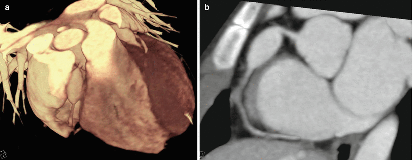
Fig. 9.1
A 6-year-old girl with Kawasaki disease presenting with non-thrombosed coronary aneurysm. (a) 3D volume-rendering CT image shows fusiform aneurysm at the right coronary artery and the left anterior descending artery. (b) Curved multiplanar reformation (cMPR) image shows non-thrombosed fusiform aneurysm at the proximal segment of the right coronary artery
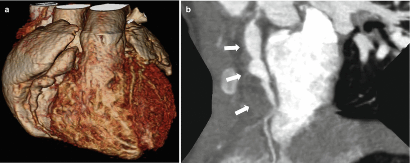
Fig. 9.2
A 3-year-old girl with Kawasaki disease presenting with thrombosed coronary aneurysm. (a) 3D volume-rendering CT image shows long segmental fusiform aneurysm at the right coronary artery. (b) On curved multiplanar reformation (cMPR) image, partial mural thrombus (arrows) was seen at the peripheral portion of huge fusiform aneurysm involving the proximal to midsegment of the right coronary artery. Actual diameter of aneurysm is much larger than that seen on 3D volume-rendering image
Takayasu arteritis
An inflammatory large-vessel vasculitis that predominantly affects young women. In early systemic phase, the involved vascular wall shows the double-ring pattern: a poorly enhanced inside ring representing mucoid or gelatinous swelling of the intima and a well-enhanced outside ring representing active medial and adventitial inflammatory change on enhanced CT (Figs. 9.3 and 9.4). In late occlusive phase, typical angiographic findings include a diffuse luminal narrowing or occlusion with/without circumferential calcification of the aorta and major branches [10, 11] (Fig. 9.5). The incidence of coronary artery involvement has been reported to be 9–10 %. Signs and symptoms result from ischemia due to arterial stenosis or occlusion. On the basis of pathological features, coronary artery lesions can be classified into the following three types: type 1 (most common), stenosis or occlusion of the coronary ostia and the proximal segments of the coronary arteries; type 2, diffuse or focal coronary arteritis, which may extend diffusely to all epicardial branches or may involve focal segments, so-called skip lesions; and type 3, coronary aneurysm [12]. Aneurysms and ectasia can also develop as a compensatory mechanism [5].

Fig. 9.3
A 49-year-old woman with active stage of Takayasu arteritis. (a) Transaxial contrast-enhanced CT image shows concentric wall thickening at the right brachiocephalic and the left subclavian arteries. (b) Transaxial contrast-enhanced CT image shows typical “double-ring” sign with poorly enhanced inner ring and well-enhanced outer ring at the ascending and descending thoracic aorta
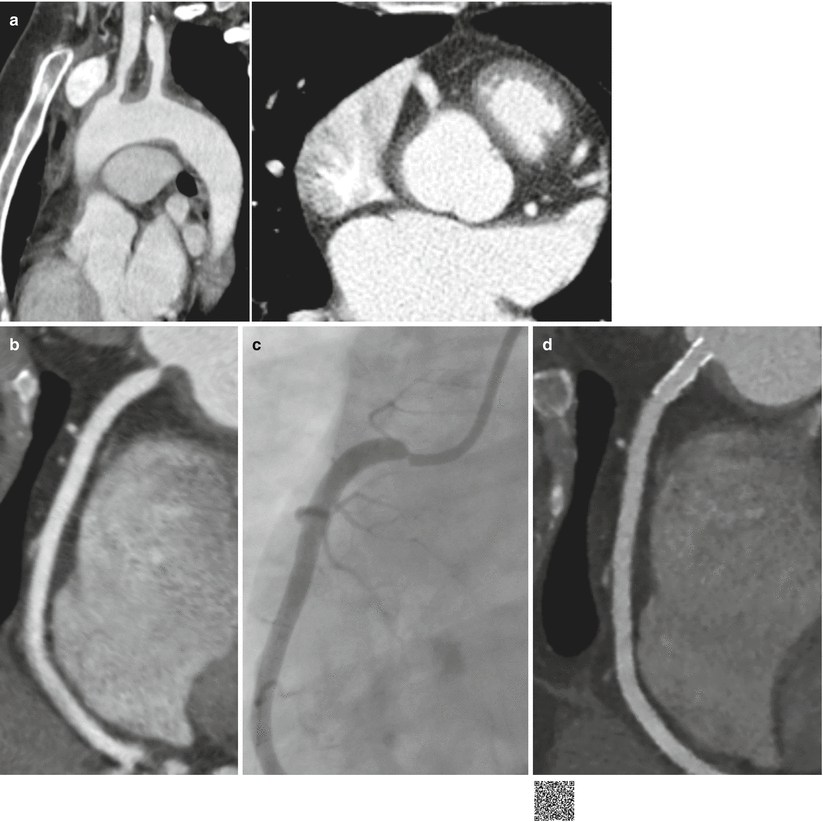
Fig. 9.4
A 29-year-old woman with Takayasu arteritis involving the right coronary artery ostium (Courtesy of Dong Hyun Yang, Asan Medical Center). (a) Oblique sagittal multiplanar reformation image shows diffuse wall thickening at the aortic arch and its branches. (b) Transaxial (b) and curved multiplanar reformation (c) images clearly demonstrate the tight luminal narrowing at the ostium of the right coronary artery by extension of inflammation presenting as wall thickening of the ascending aorta into the right coronary artery. (c) Invasive coronary angiogram shows same feature of luminal stenosis at the ostium of the right coronary artery. (d) The patient underwent subsequently stent implantation in the right coronary artery. Curved multiplanar reformation image shows excellent luminal patency of stent implanted in the right coronary artery (http://extras.springer.com/2015/978-3-642-36396-2)
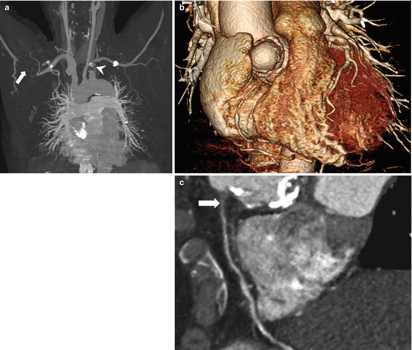
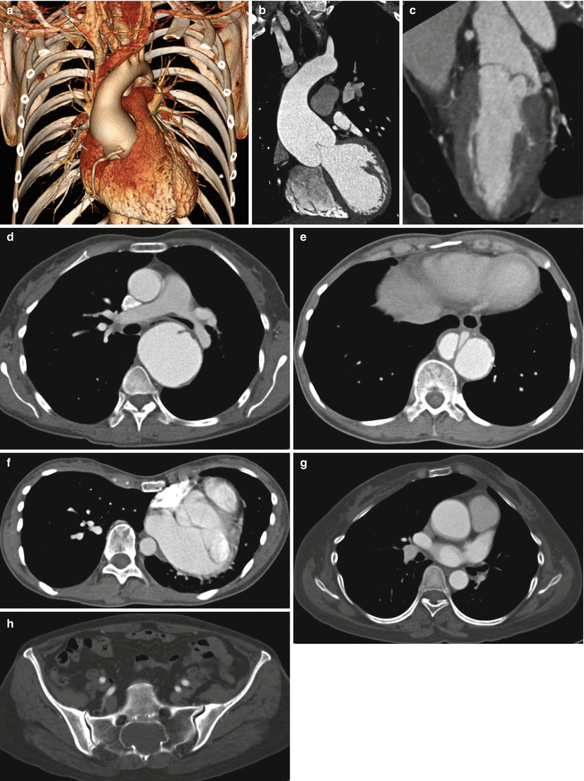
Fig. 9.5
A 55-year-old woman with Takayasu arteritis. (a) Maximum intensity projection (MIP) image shows segmental total occlusion involving the left subclavian artery (arrowhead) and the right axillary artery (arrow). (b) Three-dimensional volume-rendering image shows focal outpouching bizarre aneurysm with ring calcification at the right aortic sinus, indicating unusual manifestation of Takayasu arteritis. The right coronary artery ostium was occluded and diffuse narrowing of proximal segment was seen. (d) Curved multiplanar reformation (cMPR) image also shows the occluded right coronary artery proximal segment (arrow)
Other reported causes of inflammatory coronary vasculitis are listed in Table 9.1. Unlike other collagen vascular diseases such as polyarteritis nodosa and rheumatoid arthritis, in which coronary arteritis has been reported in 62 and 20 %, respectively, of patients who underwent autopsy, coronary vasculitis is quite uncommonly seen in systemic lupus erythematosus (SLE) [13]. Twelve cases of SLE-associated coronary aneurysms have been reported in the literatures, involving focal or diffuse and one to three coronary arteries [14]. Myocardial infarction in SLE is caused from either coronary arteritis or premature atherosclerosis. The majority of cases are secondary to atherosclerosis, which is believed to be accelerated in those treated with corticosteroids. Coronary arteritis accounts for only very few cases of myocardial infarction in patients with SLE. The representative features of coronary arteritis differentiating from atherosclerosis in SLE on the basis of changes in coronary anatomy found by angiography are reported as smooth focal lesions, aneurysmal dilatation, and an abrupt consecutive change from normal to severe obstruction of coronary arteries [13] (Fig. 9.6). The inflammation from any causes including infection may lead to in situ coronary thrombosis as well [1].
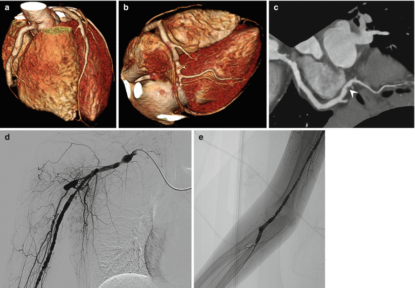
Fig. 9.6
A 22-year-old woman with systemic lupus erythematosus. (a, b) Three-dimensional volume-rendering images show diffuse dilatation (ectasia) with combined stenosis of the right coronary artery and the posterolateral branch. (c) Curved multiplanar reformation image shows focal wall thickening (arrowhead) at the site of luminal narrowing of the posterolateral branch. (d, e) Upper extremity angiograms show diffuse aneurysmal dilatation (ectasia) with multifocal combined stenosis of the brachial artery and its branches. The angiogram features look like “string of beads” appearance similar to fibromuscular dysplasia
9.2.2 Connective Tissue Diseases
Ehlers-Danlos syndrome, Marfan syndrome, and Loeys-Dietz syndrome are genetic disorders that primarily affect the soft connective tissues of multisystem (Fig. 9.7). Histologically, the aortic media shows a deficiency of elastic and muscle fibers, naming cystic medial necrosis [15]. Association of coronary artery dissection from extension of a dissection from proximal aorta has been reported in patients with connective tissue diseases such as Marfan syndrome or Ehlers-Danlos syndrome [15]. Although true aneurysm of the coronary artery in Marfan syndrome is very rare, interestingly there have been predilection for the location and timing of aneurysms; there have similar reports showing dilatation of coronary origin during follow-up after total repair of annuloaortic ectasia [16, 17] (Fig. 9.8). Coronary artery aneurysms in patients with Noonan syndrome have also been reported. The association of coronary artery aneurysm with Noonan syndrome is not well understood. Several pathologic mechanisms have been proposed, including vasculitis superimposed upon a connective tissue defect, dilatation secondary to associated myocardial hypertrophy, and persistent aneurysm after the spontaneous closure of fetal coronary artery fistula [18].
 < div class='tao-gold-member'>


Only gold members can continue reading. Log In or Register to continue
Stay updated, free articles. Join our Telegram channel
Full access? Get Clinical Tree
