Remarks
There are familial cases of anomalous origin of the coronary arteries, in which a family screening would be recommended
Anomalies of the coronary arteries can be the cause of death with different degrees of certainty:
Certain cause: anomalous origin from the pulmonary artery
Probable cause: anomalous left anterior descending or left coronary artery origin from the right aortic sinus with an interarterial course
Uncertain cause: anomalous right coronary artery origin from the left aortic sinus, anomalous left anterior descending coronary artery origin from the right aortic sinus without an interarterial course, high take-off origin, intramyocardial bridge
6.2 Congenital Anomalies
Congenital anomalies of the coronary arteries constitute the most frequent cause of sudden death (SD) from non-atheriosclerotic coronary diseases (second cause of death in athletes in the USA). In the majority of cases, SD occurs during or immediately after exercise. There are several classifications and their incidence varies depending on the source of the published study (whether based on necropsies, coronary angiographies, echocardiographies, multisection CT, cardiac MRI, etc.), although in general it can be assumed that the incidence ranges from 0.3 to 1.3 %.
The clinical spectrum and presentation is variable, from asymptomatic patients to patients presenting with angina, acute myocardial infarction and sudden death. The major concern for clinicians is to establish which type of coronary artery anomaly may lead to SD. In young people (<30 years), when the anomalous coronary artery is the dominant one or when the course is interarterial (between the aorta and pulmonary artery) or intramural, the risk of sudden death increases.
The most frequent causes are summarised in Table 6.1.

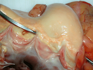
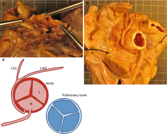
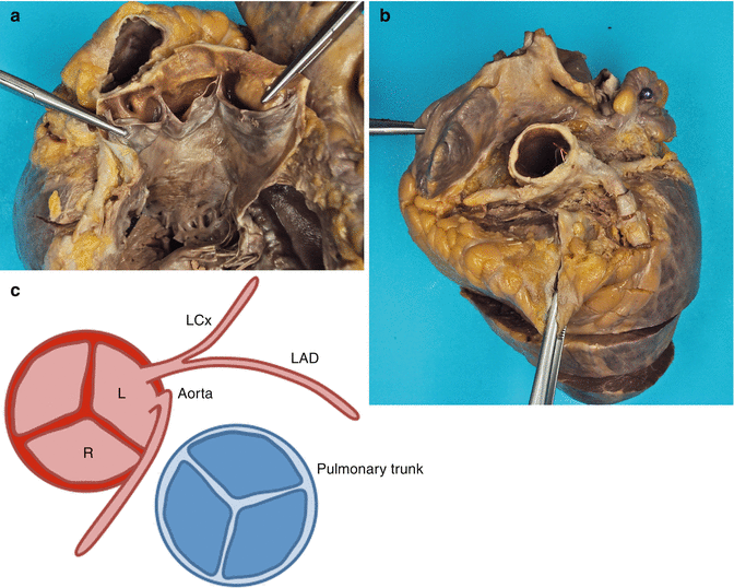
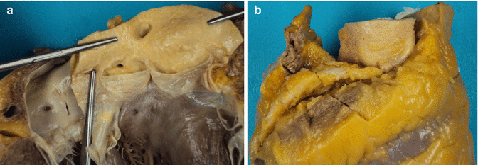
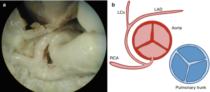
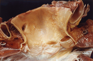
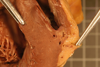
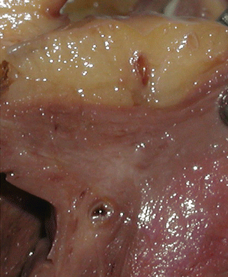
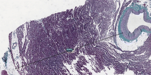
Table 6.1
Congenital anomalies most related to SD
Anomalous origin of the left coronary artery from pulmonary artery (Fig. 6.1) |
Ostium in left pulmonary sinus (most frequent) |
Dilated and tortuous right coronary artery and its branches |
Signs of chronic ischaemia and left ventricular hypertrophy are frequently found |
Anomalous ostium close to the aortic valve commissure, and frequently slit-like. Acute angle between the aorta and the coronary artery |
The course can be interarterial (towards the right in the epicardium or within the aorta adventitia), retroaortic (towards the left) or within the septum |
Signs of chronic ischaemia and left ventricular hypertrophy are frequently found |
Anomalous ostium similar to the above |
Signs of myocardial ischaemia are generally not found |
Single coronary ostium and artery (Fig. 6.6) |
Single left or right coronary artery originating the rest of the branches |
Ostium located more than 1 cm above the sinotubular junction |
Usually buttonhole-shaped with an initial intramural course |
Depth of the tunnel more than 5 mm or depth of 2–3 mm for 2–3 cm in length |
Morphology of the surrounding myocardium similar to a sphincter |
Perivascular myocardial disarray and fat degeneration |

Fig. 6.1
Anomalous origin of the left coronary artery from the pulmonary artery. An 11-year-old boy who died suddenly after a race. (a) Origin of the left coronary artery from the pulmonary artery. (b) Very dilated origin of the right coronary artery in the normal location (right aortic sinus)

Fig. 6.2
Anomalous origin of the left coronary artery from the right aortic sinus. Sudden death after prolonged exertion in a 47-year-old male. The heart showed atherosclerotic plaques in the aorta, severe coronary atherosclerosis in the proximal course of the LAD coronary artery and subendocardial scarring fibrosis in the LV. In this case, the cause of death should be attributed to the coronary atherosclerosis rather than to the congenital anomaly. However, the anomalous origin of the coronary artery may speed up the atherosclerotic process given the turbulence in the blood flow caused by the presence of acute angles in its course

Fig. 6.3
Anomalous origin of the left coronary artery from the right aortic sinus. An 8-year-old boy who suffered a cardiac arrest episode when coming off a fairground attraction. (a) Both coronary arteries arise from the right coronary sinus, the left coronary ostium is located below the right one and its calibre is smaller. (b) The right coronary artery follows its usual path, but the left coronary artery turns to the right and backwards, originating an acute take-off angle, continuing its course within the aorta’s adventitia and surrounds the posterior side of the aorta up to its bifurcation (Saldaña et al., (2009). Published with permission from Cuadernos Medicina Forense). (c) Diagram representing this anomaly

Fig. 6.4
Anomalous origin of the right coronary artery from the left aortic sinus. A 43-year-old male who died suddenly after complaining for several days of pain in the left arm. (a, b) Anomaly in the RCA that arises from the left sinus and has an intramural aortic course. (c) Diagram representing this anomaly

Fig. 6.5
High take -off and displacement to the left of the right coronary ostium. A 49-year-old male who died in hospital while undergoing biopsy of a tumoural tongue lesion. (a) Right ostium 1.5 cm above the sinotubular junction and displaced laterally to the left over the anterior commissure, at the junction between the right and the left Valsalva sinuses. (b) Detail of the high take-off of the right coronary artery with an acute angle take-off and an intramural aortic course of 2 cm (arrow) until it reaches its natural path underneath the right atrial appendage

Fig. 6.6
Common origin of both right and left coronary arteries in the right aortic sinus (single coronary artery). A 4-month-old infant male with malformation syndrome of unknown aetiology with karyotype 47XYY. Sudden death at home. (a) Single coronary ostium in the right coronary sinus from where a common trunk arises that immediately bifurcates in both left and right coronary arteries. The RCA keeps its normal course whereas the LCA surrounds the aortic arch in its posterior side and courses to reach the anterior part of the heart, where it originates the circumflex and LAD arteries. (b) Diagram representing this anomaly

Fig. 6.7
High take-off origin of the right coronary artery. A 60-year-old woman who died suddenly at home. Autopsy: hydrocephalia with subependymal gliosis and dilated cardiomyopathy (heart weight: 815 g. LV: 5 cm in diameter)

Fig. 6.8
Intramyocardial course of the left anterior descending coronary artery. A 47-year-old male who died suddenly at home after an episode of chest pain. Heart weight of 418 g with biventricular hypertrophy (RV wall thickness: 0.5 cm; LV wall thickness: 1.5 cm. The LAD coronary artery was dilated in its proximal portion and from the mesocardium, the course turns intramyocardial for 4 cm in which it tunnels through the interventricular septum at a depth of 7 mm from the epicardium (arrow), emerging again to it. Areas of fibrosis and perivascular adipose infiltration can be appreciated in the myocardium surrounding the bridge

Fig. 6.9
Intramyocardial course of the left anterior descending coronary artery. Sudden death in a 27-year-old male. The middle third portion of the LAD coronary artery dips into the myocardium, at a depth of 0.7–0.8 cm and for a length of 1.3 cm. Areas of fibrosis and perivascular adipose infiltration in the myocardium surrounding the bridge

Fig. 6.10
Histological section of a myocardial bridge. Sudden death in a 33-year-old male. Intramural course of the middle third of the left anterior descending coronary artery (myocardial bridge) of 5.065 mm depth and 20 mm length. Band of subepicardial myocardial tissue enveloping the coronary artery, causing compression of the artery during systole (Masson’s trichrome)
6.2.1 Anomalous Origin of the Coronary Arteries from the Opposite Aortic Sinus
This is the most common congenital anomaly. There are different variations according to the location of the ostium, to whether they originate from a common orifice or from two separate ones, and to the course of the anomalous coronary artery until it reaches its dependent territory. Thus, an interarterial course (between the aorta and pulmonary artery) is associated with a greater compromise in arterial blood flow and consequently with increased lethality. Some authors suggest that anomalous courses are associated with a higher presence of atherosclerotic disease.
6.2.2 Anomalous Origin of the Coronary Arteries from the Pulmonary Trunk
Although there are some instances where both coronary arteries originate from the pulmonary artery, the most frequent situation is anomalous left coronary artery from the pulmonary artery, and in 95 % of the cases from the left pulmonary sinus. Until recently, diagnosis of this anomaly in an infant was associated with high mortality, although currently the use of non-invasive imaging techniques has allowed the diagnosis of new cases in the adult population with an average age of 41 years. The right or left anterior descending coronary arteries can also arise from the pulmonary artery but the frequency is very low and these anomalies are generally asymptomatic, although in some cases they have been described associated with sudden death.
6.2.3 High Take-off Origin
This congenital anomaly is an infrequent cause of sudden death. The coronary ostia are usually located in the Valsalva sinuses although they can also be situated above them, especially the left ostium. A location of the ostium up to 2–3 mm above the sinotubular junction is considered normal. Although there is no conclusive measurement it is generally accepted that high take-off origin occurs when the distance to the Valsalva sinus is greater than 1 cm. In these cases, the initial course of the coronary artery is vertical and intramural with an acute take-off angle that hinders diastolic filling. The ostium is buttonhole-shaped.
< div class='tao-gold-member'>
Only gold members can continue reading. Log In or Register to continue
Stay updated, free articles. Join our Telegram channel

Full access? Get Clinical Tree


