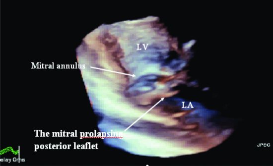
Figure 9.2 Three-dimensional echocardiography shows mitral valve prolapse. LA, left atrium; LV, left ventricle.
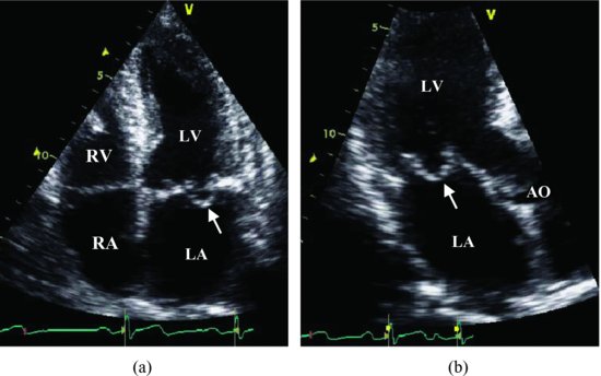
For comparison, we put another case with mitral posterior leaflet prolapse in Figure 9.3 and Videoclip 9.2.
Figure 9.3 Apical 4-chamber view shows the mitral posterior leaflet extends superior to a line connecting the annular hinge points (arrow) (a). The apical long-axis view shows the mitral posterior leaflet prolapse (b). AO, aorta; LA, left atrium; LV, left ventricle; RA, right atrium; RV, right ventricle.
Discussion
Stay updated, free articles. Join our Telegram channel

Full access? Get Clinical Tree


