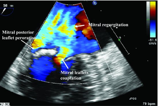
Figure 79.2 Transesophageal echocardiography image with color Doppler (systole) shows two jets of mitral regurgitation, one through mitral leaflets coaptation, another anterior direction jet from the mitral posterior leaflet perforation.
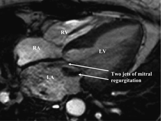
Magnetic resonance image found two jets of regurgitation consistent with TEE image (Figure 79.3, Videoclip 79.2). Culture was obtained from the incision. This grew out 1+ coagulase-negative Staphylococcus (not Staphylococcus aureus).
Figure 79.3 Magnetic resonance image illustrates two jets of regurgitation, one through mitral leaflets coaptation, another anterior direction jet from the mitral posterior leaflet perforation. LA, left atrium; LV, left ventricle; RA, right atrium; RV, right ventricle.
Discussion
Stay updated, free articles. Join our Telegram channel

Full access? Get Clinical Tree


