and Mark C. Mammel2, 3
(1)
Division of Neonatal-Perinatal Medicine, C.S. Mott Children’s Hospital University of Michigan Health System, Ann Arbor, MI, USA
(2)
Neonatal Medicine, Children’s Hospitals and Clinics of Minnesota, St. Paul, St. Paul, MN, USA
(3)
University of Minnesota, Minneapolis, MN, USA
Keywords
Endotracheal tubeTurbulent flowAir leak6.1 Endotracheal Tube Leaks
Because cuffed endotracheal tubes are not used in newborns, there is always some degree of gas leak around the endotracheal tube. Leaks can be detected graphically, using either waveform or loop analysis.
Figure 6.1 is a tidal volume (Vt) waveform demonstrating a large endotracheal tube leak. Normally, the descending portion of the waveform reaches the zero baseline at end-expiration. However, in the face of a leak, the waveform fails to reach the baseline and may have the appearance similar to a pressure waveform with positive end-expiratory pressure (PEEP).
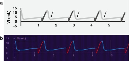
Figure 6.1
Large endotracheal tube leak (a, schematic; b, actual). The volume waveform is abnormal; it fails to reach the zero baseline (arrows)
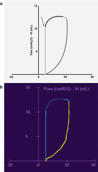
Leaks also affect the pressure-volume (P-V) and the flow-volume (F-V) loops. Fig. 6.2 shows a P-V loop from a baby with a large endotracheal tube leak. The deflation limb stops before reaching the PEEP value (the straight solid line is an artifact drawn by the monitor). Fig. 6.3 is the F-V loop from the same patient. Here the expiratory flow terminates prematurely, reaching a zero flow state long before the origin (again, the straight solid line is a monitor-generated artifact). Many ventilators are able to quantify the leak by comparing the difference between inspired and expired Vt; some will demonstrate this.