Miscellaneous Benign and Low-Grade Tumors
Allen P. Burke, M.D.
Fabio R. Tavora, M.D., Ph.D.
Hemangioma
Clinical
Cardiac hemangiomas constitute fewer than 10% of benign cardiac tumors. Approximately 200 have been reported.1 They occur in all ages with a mean in the 5th decade. Up to 25% of cardiac hemangiomas occur in infants and young children.2 Most cardiac hemangiomas are discovered incidentally. Cardiac hemangiomas may cause arrhythmias, pericardial effusions, congestive heart failure, outflow tract obstruction, and rarely sudden death.2 Extracardiac hemangiomas can occur in association with cardiac lesions, especially in the gastrointestinal tract.3
Pathologic Findings
Grossly, cardiac hemangiomas are usually mural lesions but can also be found in the subendocardial space, as dark red well-circumscribed nodules that measure from <1 to 4 cm or larger in maximum diameter. Those endocardial lesions are usually of the capillary type histologically. In contrast, the less common intramural hemangiomas that arise with the ventricular wall resemble intramuscular hemangiomas of soft tissue.3 Approximately half of these show at least focal thickened vessel walls that suggest arteriovenous malformations, and fat and fibrous tissue are not uncommon.
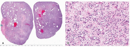 FIGURE 183.1 ▲ Cardiac hemangioma. A. Low magnification demonstrates a cellular tumor with scattered spaces (dilated vascular channels). B. Higher magnification of a cellular area shows features of capillary hemangioma. |
Endocardial-based hemangiomas of the heart are occasionally misdiagnosed as myxomas, because of their vascularity and myxoid stroma (Fig. 183.1). However, their vessels are mature and, in contrast to myxoma, are invested with actin-positive pericytes. In further distinction from myxoma, hemangiomas are calretinin negative.
Endocardial-based hemangiomas may also be confused with organized thrombus. However, organized thrombi will in some areas demonstrate typical features of entrapped blood, fibrin, and hemosiderin. Hemangiomas that infiltrate the trabeculae of the right atrium may be particularly difficult to distinguish from organized thrombus (Fig. 183.2).
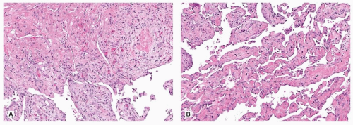 FIGURE 183.2 ▲ Cardiac hemangioma. A. The interface between the right atrial muscle (above) and tumor (below) is seen. The diagnosis is subtle in this area. B. Another area of the same tumor showing a papillary growth pattern that can occasionally cause diagnostic difficulties with angiosarcoma. |
Benign Fatty Tumors
Lipomatous Hypertrophy of the Atrial Septum
Lipomatous hypertrophy of the atrial septum (LHAS) is a tumor-like condition characterized by excessive deposition of fat in the interatrial septum and a thickness of >2 cm4,5 (Fig. 183.3A). It has been considered a form of hamartoma of immature brown fat. It has also been speculated to be part of a metabolic condition since there seems to be a strong correlation of LHAS with obesity.5 LHAS, although anatomically located within the atrial septum, has a tendency to bulge into the right atrium. For this reason, the lesions are often interpreted by computed tomography or magnetic resonance as right atrial tumors.
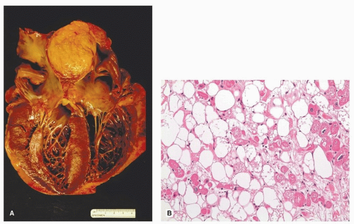 FIGURE 183.3 ▲ Lipomatous hypertrophy, atrial septum. A. Gross photograph demonstrates incidental atrial septal thickening by fat. The ventricles are dilated (dilated cardiomyopathy). B. Histologic section shows vacuolated brown fat cells and interspersed myocytes, which typically have enlarged hyperchromatic nuclei. |
Of the clinically apparent cases, the two most common signs are related to supraventricular arrhythmias and atrioventricular or right atrial inflow obstruction.4 Computed tomography and magnetic resonance can also be useful investigations for diagnosis and in planning surgery. Surgical excision of the septal fat necessitates atrial reconstruction with pericardial or Dacron patches.
Macroscopically, LHAS cannot be distinguished from normal epicardial fat. There is usually no tumor capsule.4
Stay updated, free articles. Join our Telegram channel

Full access? Get Clinical Tree


