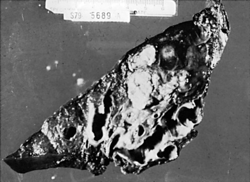
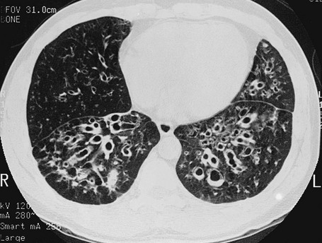
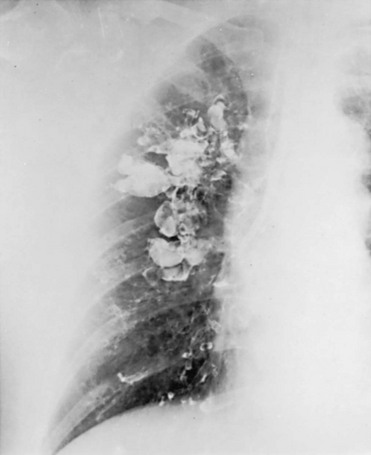
Miscellaneous Airway Diseases

7


Miscellaneous Airway Diseases


This chapter considers several disorders that affect airways, chosen because of their clinical or physiologic importance. The first of these disorders, bronchiectasis, is a disease that was much more common in the past. The availability of effective antibiotics for control of respiratory tract infections has made this problem less prevalent and diminished its clinical consequences. The second disorder, cystic fibrosis, is a genetic disease that generally manifests in childhood and is notable for the often devastating clinical consequences that ensue. Abnormalities of the upper airway (which for our purposes here includes the airway at or above the level of the trachea) are discussed briefly to acquaint the reader with the physiologic principles that allow detection of these disorders.


Bronchiectasis


Bronchiectasis is an irreversible dilation of airways caused by inflammatory destruction of airway walls. Because the most common etiologic factor is infection, which triggers the destructive inflammatory process, the extent of the bronchiectasis in a patient depends on the location and extent of the underlying infection. In some cases, bronchiectasis is localized to a specific region of the lung. In other cases, the process involves more than one area or even is diffuse, involving a large portion of both lungs.


Etiology and Pathogenesis


Infection and impairment of drainage (frequently due to obstruction) are the two underlying problems that contribute to development of dilated or bronchiectatic airways. The responsible infection(s) may be viral or bacterial. Years ago, measles and pertussis (whooping cough) pneumonia were common problems resulting in bronchiectasis. Currently, a variety of other viral and bacterial infections often are responsible; important examples are tuberculosis and Mycobacterium avium complex. At times, inflammation resulting from hypersensitivity to fungal organisms is the underlying cause, as with allergic bronchopulmonary aspergillosis. This condition, found almost exclusively in patients with clinically apparent asthma or cystic fibrosis, is characterized by colonization of airways with Aspergillus organisms and by thick mucous plugs and bronchiectasis in relatively proximal airways.


When an airway is obstructed, a superimposed infection may develop behind the obstruction, causing destruction of the airway wall and leading to bronchiectasis. Tumors, thick mucus, or foreign bodies commonly cause bronchial obstruction, resulting in bronchiectasis.


Another factor that plays a role in some patients is a defect in the ability of the airway to clear itself of, or protect itself against, bacterial pathogens (see Chapter 22). Such a defect predisposes a person to recurrent infections and eventually to airway dilation and bronchiectasis. The abnormality may involve inadequate humoral immunity and insufficient antibody production (hypogammaglobulinemia) or defective leukocyte function. Another problem that has received significant attention is dyskinetic cilia syndrome, in which ciliary dysfunction affects the ability of the ciliary blanket that lines the airway to clear bacteria and protect the airway against infection. The ciliary dysfunction is not limited to the lower airways; it also affects the nasal mucosa and, in males, may affect sperm motility and hence fertility. Pathologically, the dynein arms that are a characteristic feature of the ultrastructure of cilia are frequently absent in this disorder. One specific syndrome associated with bronchiectasis and ciliary dysfunction is Kartagener syndrome, which includes a triad of sinusitis, bronchiectasis, and situs inversus (usually discovered because of the presence of dextrocardia).


Pathology


The primary pathologic feature of bronchiectasis is evident on gross inspection of the airways, which are markedly dilated in the involved region (Fig. 7-1). Three specific patterns of dilation have been described: cylindrical (appearing as uniform widening of the involved airways), varicose (having irregularly widened airways resembling varicose veins), and saccular bronchiectasis (characterized by widening of peripheral airways in a balloonlike fashion). These terms are still used when describing radiographic patterns but are much less relevant clinically. The dilated airways are generally filled with a considerable amount of secretions that may be grossly purulent. Microscopic changes of the bronchial wall epithelium, consisting of ulceration and squamous metaplasia, are seen.


As a result of the exuberant inflammatory changes in the bronchial wall, the blood supply, provided by the bronchial arteries, is increased. The arteries enlarge and increase in number, and new anastomoses may form between the bronchial and pulmonary artery circulations. Inflammatory erosion or mechanical trauma at the site of these vascular changes is often responsible for the hemoptysis seen frequently in patients with bronchiectasis.


Coexisting disease in the remainder of the tracheobronchial tree is common. Other areas of bronchiectasis may be present, or generalized changes of chronic bronchitis may be seen (see Chapter 6).


Pathophysiology


Once the airways have become irreversibly dilated, their defense mechanisms against infection are disturbed. The normal propulsive action of cilia in the involved area is lost, even if it was intact before development of bronchiectasis. Bacteria colonize the enlarged airways, and secretions pool in the dilated sacs of patients with saccular bronchiectasis. Cough becomes much less effective at clearing secretions because of the abnormally collapsible airways. In many cases the relationship established between the colonizing bacteria and the host is relatively stable over time, but the course may be punctuated by acute exacerbations of airway infection.


Functionally, patients with a localized area of bronchiectasis are not impaired to the same extent as patients with generalized obstructive lung disease. Measurement of pulmonary function may reveal surprisingly few if any abnormalities. When seen, functional abnormalities are the result of either extensive bronchiectasis involving a large area of one or both lungs or coexistent generalized airway disease, primarily chronic bronchitis.


Clinical Features


The most prominent symptoms in patients with bronchiectasis are generally cough and copious sputum production. The sputum may be frankly purulent and tenacious, and often the profuse amount of yellow or green sputum production raises the physician’s suspicion of bronchiectasis. However, not all patients with bronchiectasis have significant sputum production. It has been estimated that approximately 10% to 20% of patients are free of copious sputum production; these patients are said to have “dry” bronchiectasis.


The other frequent symptom in patients with bronchiectasis is hemoptysis, which may be massive and life threatening. Hypertrophied bronchial arterial circulation to the involved area is responsible for this symptom in the majority of cases. Because bronchial arteries are branches of the aorta and therefore perfused at systemic blood pressure, bleeding from these vessels can be brisk. Physical examination of the patient with bronchiectasis may reveal few abnormalities, even over the area of involvement. On the other hand, the examiner may hear strikingly abnormal findings such as wheezes, crackles, or rhonchi in a localized area. Clubbing is frequently present. Although the mechanism is not clear, clubbing is thought to be associated with the chronic suppurative process.


Whether arterial blood gas values are abnormal in these patients often depends on the extent of involvement and the presence or absence of underlying chronic bronchitis. With well-localized disease, both PO2 and PCO2 may be normal. At the other extreme, patients with more severe disease may develop hypoxemia and hypercapnia. Cor pulmonale may subsequently develop.


Diagnostic Approach


The diagnosis of bronchiectasis is usually suggested by a history of copious sputum production, hemoptysis, or both. Evaluation on a macroscopic level generally includes a chest radiograph, which often reveals nonspecific abnormalities in the involved area. The radiograph may show an area of increased markings, crowded vessels, or “ring” shadows corresponding to dilated or saccular airways. However, none of the findings on the routine radiograph is considered diagnostic of bronchiectasis.


High-resolution computed tomography (HRCT) provides a definitive diagnosis and is the initial procedure used to define the presence, location, and extent of bronchiectasis (Fig. 7-2). HRCT (with sections 1-2 mm thick) provides excellent detail and is particularly useful for detecting subtle bronchiectasis. In the past, the definitive diagnosis depended on bronchography, a radiographic procedure in which an inhaled opaque contrast material was used to outline part of the tracheobronchial tree (Fig. 7-3). This procedure is uncomfortable, can induce bronchospasm, and is not performed today.


Evaluation on a microscopic level is not particularly helpful for patients with presumed bronchiectasis, except for examination of the sputum for microorganisms, particularly during an acute exacerbation of the disease. Patients with bronchiectasis frequently become colonized and infected with Pseudomonas aeruginosa, and the finding of this otherwise relatively unusual pathogen may be a clue to the presence of underlying bronchiectasis. The findings on functional evaluation were discussed in the sections on pathophysiology and clinical features.


Treatment


The three major aspects of treatment of bronchiectasis are antibiotics, bronchopulmonary drainage (clearance of airway secretions), and bronchodilators. Antibiotics are used in various ways. Some patients are treated only when the quantity or appearance of the sputum clearly changes. Other patients, especially those who have severe disease and frequent exacerbations, are given a regimen of intermittent or even continuous antibiotics in an attempt to control chronic infection. Oral agents such as amoxicillin and trimethoprim-sulfamethoxazole, which are effective against many strains of Streptococcus pneumoniae and Haemophilus influenzae, are often used in patients with bronchiectasis. Inhaled tobramycin is sometimes used prophylactically to diminish the growth of gram-negative organisms. When these patients are infected with Pseudomonas organisms, treatment is generally more difficult. Oral fluoroquinolones such as ciprofloxacin have become useful therapy for Pseudomonas infection as an alternative to parenteral antibiotics, but secondary development of resistance to this class of antibiotics is common. Infection with M. avium complex requires prolonged therapy with multiple drugs (see Chapter 24

Only gold members can continue reading. Log In or Register to continue

Stay updated, free articles. Join our Telegram channel

Jun 12, 2016 | Posted by in RESPIRATORY | Comments Off on Miscellaneous Airway Diseases

Full access? Get Clinical Tree

Get Clinical Tree app for offline access