CHAPTER 30 Minimally Invasive Surgery for Atrial Fibrillation
Approach to Video-Assisted Thoracic Surgery for Atrial Fibrillation
Key Points
♦ Patients who choose a minimally invasive surgical approach to correcting atrial fibrillation usually are otherwise in good health. There is no room for error, and the mortality rate must be zero.
♦ Our bilateral VATS technique is designed to allow cardiac surgeons to achieve atrial fibrillation cures safely but without mastering totally thoracoscopic skills.
Video-Assisted Surgery for Atrial Fibrillation
Step 1. Setup
♦ Use a double-lumen endotracheal tube for selective lung ventilation, and place the central line, arterial line, and external defibrillator pads in the appropriate vector.
Step 2. Patient Positioning and Port Placement
♦ Place the patient in the left lateral decubitus position at 45 to 60 degrees, with the right arm on an LPS Arm Support (Allen Medical, Acton, Maine).
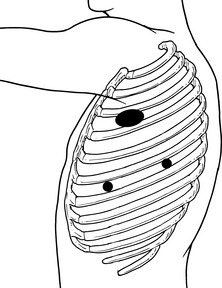
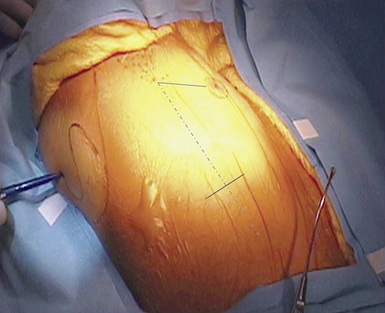
♦ Document the external anatomy after reviewing the internal anatomy using the chest radiograph. Outline the scapula, mid-axillary line, and a line from the xyphoid posteriorly (Figure 30-1).
♦ First port placement
 Place the first port in the sixth or seventh intercostal space in the mid-axillary line where the xyphoid line and mid-axillary lines cross.
 Place the first port in the sixth or seventh intercostal space in the mid-axillary line where the xyphoid line and mid-axillary lines cross.
♦ Large working port placement
 Make a 4- to 6-cm working incision from the auscultatory triangle in the third or fourth intercostal space, and carry it anteriorly.
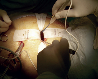
 For the muscle-sparing technique, if the third intercostal space is used, only the intercostal muscles need to be divided. If the fourth intercostal space is used, divide the serratus anterior in the direction of the muscle fibers. Avoid cutting the pectoralis muscle by retracting it anteriorly (Figure 30-3).
 Use caution posteriorly to retract the fat pad containing any neurovascular bundle with a finger when carrying the incision over the axillary contents.
 Make a 4- to 6-cm working incision from the auscultatory triangle in the third or fourth intercostal space, and carry it anteriorly.
 For the muscle-sparing technique, if the third intercostal space is used, only the intercostal muscles need to be divided. If the fourth intercostal space is used, divide the serratus anterior in the direction of the muscle fibers. Avoid cutting the pectoralis muscle by retracting it anteriorly (Figure 30-3).
 Use caution posteriorly to retract the fat pad containing any neurovascular bundle with a finger when carrying the incision over the axillary contents.


