Fig. 3.1
High parasternal short-axis image at the level of ascending aorta demonstrating a large proximal AP window
Cardiac catheterisation is reserved for those patients with suspected pulmonary vascular disease in order to assess reversibility of the pulmonary vascular resistance and whether surgery is appropriate. Cardiac MRI scan can be a useful adjunctive investigation.
Imaging Pitfalls
It is common to see echo “dropout” in the blood vessel walls where AP windows can exist; this can make assessment by 2D imaging difficult. In addition, as the pulmonary vascular resistance is high in the newborn period, little flow across the defect may be observed on colour-flow Doppler. This might explain why the diagnosis is often missed. As in our case, it is easy to attribute the clinical findings to an associated patent arterial duct or ventricular septal defect (Fig. 3.2). In the absence of a shunt, if left heart volume overload is suspected, an unusual shunt such as an AP window must be actively ruled out.
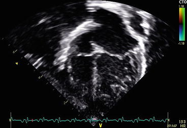
Fig. 3.2
Apical four-chamber view showing dilated left atrium and left ventricle secondary to the large left to right shunt caused by the AP window. Note the prominent pulmonary veins which are secondary to the significantly increased pulmonary blood flow
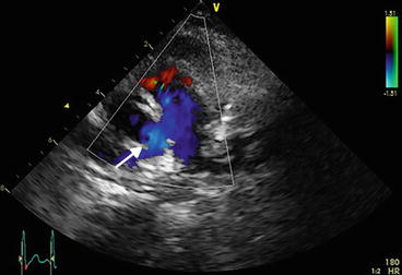
It is also easy to mis-diagnose a patient as having an AP window when the lesion is not present because of the problem of “dropout.” Figure 3.3 shows the parasternal short-axis view of a 9-month-old child who presented with mild tachypnoea and failure to thrive. He had a pansystolic murmur and enlarged liver. Echocardiogram demonstrated a moderate-sized doubly committed subarterial ventricular septal defect with left heart enlargement. In addition, the echo appearances were suspicious of a distal AP window (see Fig. 3.3). The child underwent VSD closure under cardiopulmonary bypass, but direct inspection by the surgeons ruled out a distal AP window. The volume overload was secondary to the VSD alone.
 < div class='tao-gold-member'>


Only gold members can continue reading. Log In or Register to continue
Stay updated, free articles. Join our Telegram channel
Full access? Get Clinical Tree
