Fig. 7.1
Marcello Malpighi (1628–1694)
Malpighi studied philosophy for a few years but in 1653 he turned his attention to anatomy at the University of Bologna, and this was the beginning of an extraordinarily productive career in this science. In 1656 he was invited to be professor of theoretical medicine at the University of Pisa where he began a long friendship with Giovanni Borelli (1608–1679). This man was an eminent mathematician and naturalist who was active in the Accademia del Cimento, one of the earliest scientific societies, and Malpighi became a member. However in 1659 Malpighi decided to return to Bologna and the subsequent 2 years were very productive with extensive discoveries about the lung. He moved again in 1662 to a professorship in medicine at the University of Messina, Sicily, and in fact throughout his life he spent periods at both Pisa and Messina although he always regarded Bologna as his home. His time in Messina also was very productive but he returned to Bologna in 1667.
In 1668 Malpighi received a letter from the Royal Society in London inviting him to send manuscripts to this prestigious group. Malpighi was honored by the invitation and as a result he carried out a classical study on the anatomy of the silkworm. He sent the manuscript to the Society which published it in 1669 [7] and made him a member. He maintained strong links with that group throughout the rest of his life and most of his anatomical studies were published by the Society. The admiration was mutual. On one occasion the secretary wrote to him “… to no one … observing the structure of the human body does Nature seem to have revealed her secrets as fully as to her beloved Malpighi… So too our Royal Society embraces no one with greater affection” [1].
Unhappily Malpighi was involved in a number of bitter disputes about his scientific discoveries throughout his life and he also suffered from ill health in his later years. In 1684 there was a disastrous fire at his home which destroyed many of his manuscripts and much of his equipment. In 1691 he was invited to Rome by Pope Innocent XII to be the Pope’s personal physician which was a high honor. Malpighi died of a stroke in 1694 and is buried in the church of Santi Gregorio e Siro in Bologna where there is a memorial.
Malpighi was an extraordinarily productive scientist. As we shall see he was the first person to describe the pulmonary capillaries and the alveoli. In addition he was the first to describe the anatomical basis of insect respiration as a result of his studies on the silkworm. Many of his discoveries initially bore his name and some still do. He discovered the renal glomeruli (initially called the Malpighian corpuscles), the Malpighian corpuscles in the spleen, the Malpighi layer in the skin and the Malpighian tubules in the excretory system of insects. He then went on to make extensive botanical studies, and finally did such extensive work on morphogenesis that he is sometimes called the father of embryology based on his work on the chicken embryo.
7.2 Discovery of the Pulmonary Capillaries
Much of Malpighi’s research was made possible by the recent invention of the compound microscope. Magnifying spectacles using one lens go back a long way and were in use in the thirteenth century. However the compound microscope, that is one with both an objective and an eyepiece lens, appeared much later. Some authorities attribute the invention to Hans and Zacharias Janssen in the Netherlands at the end of the sixteenth century. Galileo developed a compound microscope in 1609 but it was some time before this was exploited for scientific research. Certainly Malpighi was one of the first to use a compound microscope in 1660, and Robert Hooke’s Micrographia published in 1665 contained beautiful illustrations. Nehemiah Grew (1641–1712) and Antonie van Leeuwenhoek (1632–1723) made further important advances. However many of Malpighi’s discoveries were apparently made using a single magnifying lens
Malpighi’s historic description of the pulmonary capillaries was made in his Second Epistle to Borelli published in 1661 with the title De Pulmonibus [5]. Early in this letter Malpighi beautifully described how he came to use the frog for his dissections. He first studied sheep and other mammals but in spite of enormous efforts the results were disappointing. In fact Malpighi frequently emphasized the tediousness and inadequate results of most of his dissections.
However when he eventually used the frog he was jubilant and in a striking passage referred to the frog as the “microscope of nature”. By this he meant that he was able to visualize with a relatively small magnification, features as minute as the capillary network that had eluded him in mammals because the structures were so small that they could not be seen under his microscope. He went on to say that nature is accustomed “to undertake its great works only after a series of attempts at lower levels, and to outline in imperfect animals the plan of perfect animals”. In other words he is alluding to the fact that, as he sees it, evolution has tried out its advances in “imperfect animals” by which he means frogs before using the same structures in a more advanced form in the so-called “perfect animals” by which he means mammals. He goes on “For Nature is accustomed to rehearse with certain large, perhaps baser, and all classes of wild [animals], and to place in the imperfect the rudiments of the perfect animals”.
A little later Malpighi makes a droll statement about the amount of labor the work has taken him. “For the unloosing of these knots [that is elucidating these problems] I have destroyed almost the whole race of frogs, which does not happen in the savage Batrachomyomachia of Homer”. Here he is referring to the imaginary fierce battle between frogs and mice which is a parody of the Iliad and which, incidentally, was probably not written by Homer.
Now turning to Malpighi’s actual observations, after his abortive efforts on mammals such as sheep he first looked at the living lung in the frog. However although he could clearly see the blood moving rapidly through small arteries, he could not determine what eventually happened to it. He then dried the lung of a frog and wrote as follows, “I could not extend the power of the eye any further in the living animal, hence I believe that the mass of blood poured into an empty space and was re-collected by the outgoing vessels and the structure of their walls… however, the dried lung of a frog resolved my doubts. In a very small portion of it … there may be seen, with a perfect glass no broader than the eye, the points which are called “Sagrino” [dark spots on the surface of the lung of a frog] forming the membrane, but mixed with looped vessels. So great is the branching of these vessels, after they extend out hither and thither from the vein and artery, that no larger system of vessels will be served, but a network appears, formed by the offshoots of the two vessels. This network not only occupies the whole floor [of the airspace] but is extended to the walls and adheres to the outgoing vessel, just as I could observe more abundantly, but with greater difficulty in the oblong lung of a tortoise, which is likewise membranous and translucent. Here it lies revealed to the senses that, as the blood passes out through these twisting divided vessels, it is not poured into spaces, but is always passed through tubules and is distributed by the many windings of the vessels” (Translation from [12]).
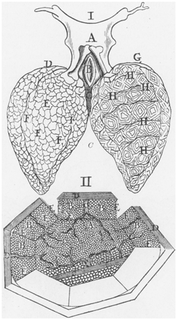
The two letters to Borelli contained two illustrations shown in Figs. 7.2 and 7.3. Malpighi’s drawing of the capillary mesh is shown in the bottom part of Fig. 7.2. This depicts one of the vesicles (alveoli) with its base and various sides that has been opened up so that the dense network of capillaries can be seen in all the walls. The upper part of Fig. 7.2 shows the two lungs of the frog. On the left we can see the alveoli and on the right is another depiction of the capillaries.


Fig. 7.2
Malpighi’s drawing of the pulmonary capillaries and alveoli. I Shows the two lungs with the alveoli on the left and the capillaries on the right. II Shows the pulmonary capillaries in a diagram of an alveolus which has been opened up. (From [5])

Fig. 7.3
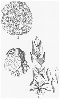
Malpighi’s drawing of the alveoli. I and II Show two different views of alveoli. III Diagram showing the branching of the trachea ending up in the alveoli. (From [5])
Malpighi’s discovery of the pulmonary capillaries was momentous. In fact it was the first description of capillaries in any circulation. Harvey in De motu cordis in 1628 had supposed, as had Galen and Columbus before him, that the blood found its way from the right ventricle through the parenchyma of the lungs into the pulmonary vein and left ventricle. However Harvey could not see the capillary vessels but called them “pulmonum caecas porositates et vasorum eorum oscilla”, that is “the invisible porosity of the lungs and the minute cavities of their vessels”. It was Malpighi’s great triumph that he was the first person to see and describe them.
7.3 Discovery of the Alveoli
Malpighi’s description of the alveoli may not have been quite so momentous as his discovery of the capillaries, but in fact it completely changed perceptions of the structure of the lung. Prior to his time the structure and function of the lung were a mystery. Vesalius following the writings of Galen held that the lungs had been formed from solidified bloody foam. Harvey compared the substance of the lung with that of the kidney or liver and argued that one of its principal purposes was to cool the animal.
Malpighi refers to these earlier notions near the beginning of his Epistle I of “De Pulmonibus” addressed to Borelli. He stated “The substance of the lungs is commonly supposed to be fleshy because it owes its origin to the blood, and it is believed to be not unlike the liver or the spleen …”. (This and the subsequent quotations from Malpighi are from the translation by Young [14].) But then in the same paragraph Malpighi goes on to drop his bombshell. “By diligent investigation I have found the whole mass of the lungs, with the vessels going out of it attached, to be an aggregate of very light and very thin membranes, which, tense and sinuous, form an almost infinite number of orbicular vesicles and cavities, such as we see in the honey-comb alveoli of bees, formed of wax spread out into partitions. These [vesicles and cavities] have situations and connection as if there is an entrance into them from the trachea, directly from the one into the other; and at last they end in the containing membrane”. Malpighi goes on to refer to one of the illustrations at the end of the Epistles which is shown in Fig. 7.3. In referring to the left-hand part of the figure he states that “with greatest diligence I have been able to make out, those membranous vesicles seem to be formed out of the endings of the trachea, which goes away at the extremities and sides into ampulus cavities”. Parts I and II of the figure show the vesicles (alveoli) and Part III is a diagrammatic representation of the final branching of the prolongations of the trachea. He adds “Seeing that the air which rushes from the trachea into the lungs requires a continuous path for easy and rapid ingress and egress, whence possible this internal tunic of the trachea, ends in sinuses and vesicles, makes a mass of vesicles like an imperfect sponge so to speak”. Although the illustrations are from the frog lung, Malpighi also refers to somewhat similar observations in a dog’s lung.
< div class='tao-gold-member'>
Only gold members can continue reading. Log In or Register to continue
Stay updated, free articles. Join our Telegram channel

Full access? Get Clinical Tree


