Fig. 23.1
Patient with a large venous malformation (VM) of the left hemiface. (a) B-mode ultrasound showing the VM as an infiltrating, hypoechoic, and compressible mass. (b, c) MRI examination: T2-weighted short TI inversion recovery (STIR) sequences acquired in coronal (b) and axial planes (c). The VM appears with a bright hypersignal, and its extension is easily delineated in adjacent soft tissues. (d) MRI examination: T1-weighted axial volumetric interpolated breath-hold examination (VIBE) MRI acquisition after gadolinium injection (MultiHance) and subtraction with baseline acquisition. The perfused area of the VM is well delineated. Several areas without perfusion are seen corresponding to phleboliths or thrombosed portions of the VM
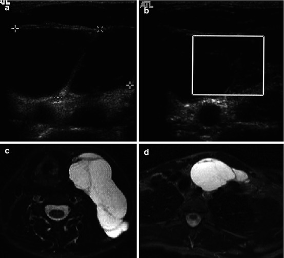

Fig. 23.2
Patient with a voluminous macrocystic lymphangioma of the left portion of the neck. (a) B-mode ultrasound showing multiple cysts with septations. (b) Color Doppler study showing a vessel with high-resistance flow in a septa. (c, d) MRI examination: T2-weighted axial (STIR) acquisitions showing the presence of fluid compatible with a macrocystic lymphatic malformation. (e) MRI examination: T1-weighted coronal acquisition after gadolinium injection showing contrast enhancement in the septations
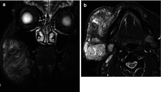
Fig. 23.3
Patient with a large microcystic lymphangioma of the right hemiface. (a, b) MRI examination: T2-weighted coronal and axial acquisitions showing a hyperintense soft tissue mass made of tiny microcysts infiltrating the cheek and masticator space
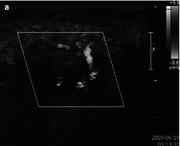
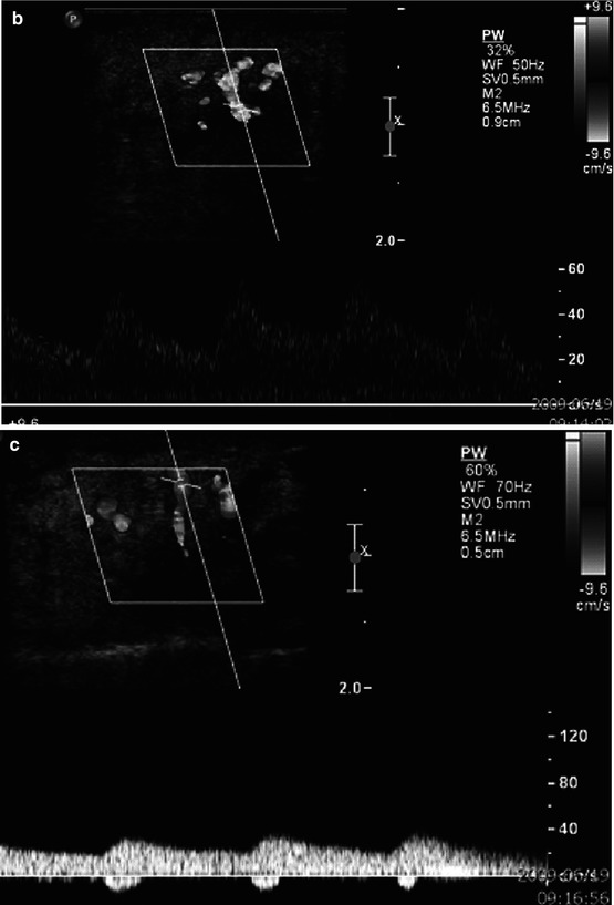
Fig. 23.4


Patient with an AVM of the lip. (a) Color Doppler ultrasound showing a high-flow malformation. (b) Pulsed Doppler ultrasound showing a high diastolic flow in the feeding artery characteristic of an arteriovenous shunt. (c) Pulsed Doppler ultrasound of a draining vein showing a systolic modulation of the flow confirming the presence of an arteriovenous shunt
Stay updated, free articles. Join our Telegram channel
Full access? Get Clinical Tree
