Management of Unusual Problems Encountered during Procedures that Require the Use of Cardiopulmonary Bypass


Management of Unusual Problems Encountered during Procedures that Require the Use of Cardiopulmonary Bypass


Mark Kurusz

Noel L. Mills

Richard F. Davis


Some uncommon preexisting clinical conditions and similarly infrequently occurring problems with the operation and/or function of the cardiopulmonary bypass (CPB) circuit can jeopardize patient safety during CPB, and may lead to adverse clinical outcomes. Given that many such occurrences present with relatively low frequency, the clinical experience of any single surgical team member or even the combined clinical experiences of several team members may be inadequate to rapidly implement an appropriate plan of management for such problems. This chapter examines several such clinical conditions and occurrences. Admittedly, some of these may stretch the definition of “unusual” and, while not representative of the day-to-day “routine” practice, should be expected to occur with a moderate frequency depending on the nature of one’s clinical practice. Some of these conditions may be encountered preoperatively, allowing time to develop a carefully considered management strategy. In contrast, some of these events may occur unexpectedly during a procedure, which demands rapid implementation of predetermined management strategies by one or more team members to prevent potentially significant morbidity or even mortality. Suggested management strategies are derived from the published literature and are referenced as appropriate, and some of the suggested strategies are derived from the authors’ collective clinical experiences.


CONDITIONS IDENTIFIABLE BEFORE CARDIOPULMONARY BYPASS


Severe Ascending Aortic Atherosclerosis

The severely atherosclerotic ascending aorta can present problems during cannulation for CPB, application of clamps, delivery of cardioplegia, construction of proximal anastomoses for coronary artery bypass grafts, or valve replacement or repair. A large multicenter study identified the presence of proximal aortic atherosclerosis as the strongest predictor of perioperative stroke (1), which is consistent with embolization of atherosclerotic debris liberated by surgical manipulation of the aorta as being perhaps the most common cause of strokes after CPB. Risk factors for ascending aortic atherosclerosis include: significant carotid, abdominal aortic, and left main coronary artery atherosclerosis; aortic wall irregularity on ascending aortic angiogram; adhesions between the ascending aorta and its adventitia; pale appearance of the ascending aorta; and minimal bleeding of an aortic stab wound (2). The presence of severe ascending aortic calcific atherosclerosis will most likely be known from preoperative imaging studies such as chest computed tomography (CT) or magnetic resonance imaging (MRI) scans, angiography or ultrasound, so that an appropriate surgical management strategy can be developed in advance of the procedure. In recent years, the use of intraoperative ultrasound, transesophageal echocardiography (TEE), or epiaortic scans has become almost universal in major cardiac surgical centers. These two techniques are complementary because of the frequent inability to visualize the upper ascending aorta with TEE due to the interposition of the tracheobronchial structures (air) between the TEE probe in the esophagus and the ascending aorta. This problem is avoided with epiaortic scanning. Visualization of Grade 4 or 5 plaques in or closely adjacent to normal aortotomy sites should prompt the use of alternative sites or surgical techniques. Cannulations via the innominate artery, the femoral artery, or the lesser curvature of the aortic arch are examples of use of alternative sites. Surgical techniques designed to minimize or avoid aortic clamping should be considered.

Careful surgical palpation of the aorta prior to selecting cannulation and other aortotomy sites may facilitate identification of less diseased sites for insertion of the arterial perfusion cannula or proximal anastomoses. Such palpation may be facilitated before CPB by inducing a brief period of hypotension with venous inflow occlusion, that is, briefly occluding the superior and inferior venae cavae to rapidly decrease cardiac ejection and arterial pressure. With lower pressure in the aorta, gentle palpation can be used to assess the quality of the aortic wall to determine the location of less diseased sites for cannulation and placement of the aortic cross-clamp. Despite these techniques, numerous reports published over the last two decades have consistently found intraoperative ultrasound imaging to be more sensitive than manual palpation for identification of atherosclerotic plaques.

In severely diseased aortas, application of an aortic crossclamp may not be deemed feasible because of increased risk
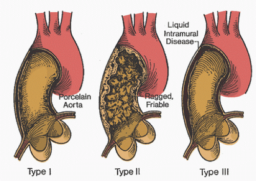
of dislodgment of atherosclerotic debris or aortic dissection (Fig. 24.1). The so-called no-touch technique has evolved to better manage these patients (2). It relies on the internal mammary artery or gastroepiploic artery as conduits for coronary bypass grafts. In some patients requiring extensive revascularization, saphenous vein grafts can be anastomosed to the internal mammary artery. In no-touch cases, a femoral artery, axillary artery, or the underside of the distal aortic arch is used as the arterial cannulation site (2,3,4). Alternatively, either a long-arch cannula can be inserted distal to the left subclavian artery or a diffusion-tipped cannula can be used (5) to better disperse the flow jet at the CPB cannula tip. However, these cannulation techniques still present some risk of stroke hazard from insertion of the cannula in the severely diseased aorta. Cannulating from the left ventricular apex with the cannula advanced through the aortic valve has also been described (6).

Rather than using conventional approaches to cardioplegia delivery, the surgeon may instead rely on electrical fibrillation or administration of a β-adrenergic blocker such as esmolol to slow the heart during distal anastomoses. In these cases, left ventricular venting may be avoided. However, if significant aortic insufficiency is present, conventional venting methods should be used, which then requires needle vent placement in the ascending aorta for de-airing before weaning from CPB and which carries some degree of hazard in the diseased aorta. An endoaortic clamp, as designed for minimally invasive cardiac surgery, may be used to isolate the heart from CPB systemic flow, deliver cardioplegic solution, and vent the aortic root (7). However, in the presence of severe atherosclerosis, this approach does not necessarily decrease the risk of dislodging atherosclerotic debris. An aortic filtration system (8) that uses an umbrella screen inserted through a modified 24F arterial cannula before aortic cross-clamp removal has been shown to capture particulate debris that presumably would have otherwise embolized systemically.


FIGURE 24.1. Type 1, type II, and type III ascending aortic atherosclerosis. No clamp is safe on these types of ascending aortic disease. Type I: Circumferential ascending aortic calcification, which may be easily diagnosed preoperatively on the angiogram. Palpation of the ascending aorta at operation reveals firm calcification. Embolization or aortic injury that may be difficult to repair may result if the aorta is clamped. Type II: This pattern may be diagnosed preoperatively by noting an irregularity of the normally smooth lining of the ascending aorta on the left ventricular angiogram or aortic root injection. Visualization of the ascending aorta is now considered a mandatory part of workup before coronary artery bypass graft. Type III: Intraluminal liquid debris is the most elusive of the three patterns to diagnose before clamping the aorta. A pale appearance of the aorta or adherence of the adventitia to the ascending aorta may be the only diagnostic clues. Operative echocardiography will reveal a thickened ascending aorta that will liberate liquid debris if a cross-clamp or partial occlusion clamp is applied. (From Mills NL, Everson CT. Atherosclerosis of the ascending aorta and coronary artery bypass: pathology, clinical correlates, and operative management. J Thorac Cardiovasc Surg 1991;102:546-553, with permission.)

In summary, cannulation of the severely diseased ascending aorta may be associated with significant morbidity after CPB. Modification of conventional CPB cannulation, cardioplegia, and venting techniques may reduce the incidence of stroke in this challenging patient population. Newer diagnostic measurements such as release of S-100b, a marker of cerebral ischemia (9), and novel perfusion cannulation techniques under development may lead to further reductions in neurologic injury in the patient with diffuse atherosclerosis needing cardiac surgery.


Hematologic Problems


Cryoproteins

Cold agglutinins are serum antibodies that become active at decreased blood temperature and produce agglutination of red blood cells. In some cases, blood cell agglutination may involve complement fixation and lead to significant hemolysis. These antibodies are most commonly directed against antigens on the red blood cells but can also be nonspecific. One very important and most clinically relevant characteristic of cold agglutinins is termed the thermal amplitude, which is the temperature below which the antibodies become activated. As temperature drops below this threshold, antibody activity increases exponentially. In general, this activity reverses as rewarming occurs (10). Another important parameter of cold agglutinins is termed the titer, which is the concentration of the cold agglutinin expressed as the dilution factor beyond which the agglutination does not occur. A 1:1 titer indicated a relatively low concentration of the cold agglutinin because a 1:1 dilution is sufficient to eliminate the agglutination, while a 1:128 titer indicated a much higher concentration of the cold agglutinin. Clearly, higher cold agglutinin titers (concentrations) are more clinically significant than low titers. There is no widely accepted definition for high versus low titer; however, Lee et al. (11) suggested titers less than 1:32 as being low and those greater than 1:128 as being high.

The complement system is activated during CPB, especially so during rewarming from hypothermia. Red blood cells agglutinated by cold agglutinins may fix activated complement and
undergo hemolysis, which can be severe, depending largely on the thermal amplitude and titer of the cold agglutinin. For hemolysis to occur, the cold agglutinin and complement activities must overlap. That is, the temperature must be low enough for the cold agglutinins to activate but warm enough for a complement fixation to occur.

Aside from during hypothermic bypass, cold agglutinins seldom produce symptoms because activation most often occurs at temperatures well below the usual range of body temperature. With cold exposure, clinical signs may include acrocyanosis of digits, tip of the nose, or ears from agglutination-induced ischemia. Most commonly, immediate warming of the affected areas reverses the agglutination and thus the ischemia. A patient with prolonged hypothermic CPB would be at risk for multi-organ damage from prolonged vascular occlusion (12). This is an uncommon but potentially catastrophic consequence of failure to recognize and treat the presence of cold agglutinins.

When patients undergo screening for cold agglutinins, a diagnosis can be easily made based on laboratory test results. If screening at 4 °C is negative, no further screening is needed. If the screen is positive at 4°C, the thermal amplitude should be determined and the titer determined for each temperature at which the screen was positive. This will give more precise information for dealing with the cold agglutinin antibodies.

In patients who are not initially screened for cold agglutinins, a diagnosis may be made by astute observation. During hypothermic CPB, agglutination within the vessels may be noted, particularly if the surgeon is wearing magnifying loops. Hemolysis manifested by hemoglobinuria is most often recognized by pink or red-tinged urine. The latter observation, however, still sometimes occurs even in the absence of cryoagglutination. If blood cardioplegia is used, the perfusionist may note agglutination in the cardioplegia delivery system as the blood is cooled (13,14). In addition, immediate agglutination of blood in a syringe during phlebotomy may indicate the presence of cold agglutinins. Agglutination also can be confirmed visually by immersing a test tube of blood into an ice slush solution and observing cell clumping on the side of the test tube that often disappears when the tube is warmed. Many cold agglutinins will present during a routine crossmatch done at room temperature. Any of the above findings suggests the presence of clinically significant cold agglutinins, and steps should be taken to prevent adverse reactions during CPB (13,15,16).

Both monoclonal and polyclonal cold agglutinins exist. The monoclonal types usually associated with lymphoreticular neoplasms are generally irreversible. The polyclonal antibodies are often associated with acute infectious diseases such as mycoplasma, infectious mononucleosis, or cytomegalovirus (12). Production of polyclonal cold agglutinins is typically transient and may remit spontaneously in weeks, but when present it may be associated with acute life-threatening intravascular hemolysis (12). Leach et al. (15) developed a comparison of clinically significant and insignificant cold agglutinins (Table 24.1). An important point in assessing the need to screen for cold agglutinins is that a failure to screen may lead to an adverse outcome, such as myocardial infarction, stroke, or acute renal failure. Without knowledge of the presence of cold agglutinins, these adverse outcomes may be attributed to another cause. For example, hemolysis may be attributed to mechanical trauma to blood from CPB (17). However, mechanical trauma to blood may be a more important source of clinically significant hemolysis than cold-reacting autoantibodies when the thermal amplitude is less than 22°C.


TABLE 24.1. Characteristics of cold agglutinins


Clinically significanta


Clinically insignificantb


  1. Lytic (cause hemolysis)


  2. Active in saline at 20°C


  3. Almost always IgM antibodies


  4. Wide thermal range (4°C-32°C)


  5. Bind complement


  6. Agglutination irreversible


  7. ↑ agglutination in albumin (30°C)


  1. Nonlytic (reversible red blood cell agglutination)


  2. Peak activity 0°C-4°C


  3. Seldom IgM antibodies (IgG, IgA, or non-Ig)


  4. Low thermal range 0°C-6°C


  5. Seldom bind complement


  6. Agglutination reversible


  7. No enhanced agglutination in albumin


a These are commonly associated with chronic cold hemagglutinin disease, neoplasm of lymphoid origin, and mycoplasma pneumonia.

b These are commonly associated with viral infections (e.g., cytomegalovirus infections and mononucleosis).


Adapted from Leach AB, Van Hasset GL, Edwards JC. Cold agglutinins and deep hypothermia. Anaesthesia 1983;38:140-143.


Treatment of cold agglutinin disease during CPB essentially consists of prevention of complement activation and ultimately of agglutination or hemolysis. Treatment is based on the etiology and the severity of the problem. In a mild case of cold agglutinins (i.e., a case in which there is a very low thermal amplitude, such as 4°C) and/or a low titer of antibodies, minor or no changes in surgical or CPB techniques and, in some cases, treatment with corticosteroids to avoid hemolysis have been advised (18). For patients with low-titer nonspecific antibodies (and only these patients), Moore et al. (10) concluded that hypothermic CPB can be performed on these patients without increased risks of hemolytic or agglutination crises; further, minor degrees of hemolysis occur in all patients during hypothermic bypass, especially during the rewarming phase, whether or not cold agglutinins are present.

In a case with clinically significant cold agglutinins (i.e., high thermal amplitude, high titer, or clinical symptoms), a number of changes in surgical technique have resulted in successful surgery using CPB. In the case of clinically significant cold agglutinins caused by acute infection (e.g., a recent viral illness), elective cardiac surgery should be postponed for
several weeks, by which time the antibody may have disappeared (17). If the urgency of surgery precludes that approach, the most sensible approach is to use either normothermia or mild hypothermia using blood temperatures continuously maintained above the thermal amplitude to avoid the active temperature range of agglutination (17,19,20,21,22,23). Hence, the presence of cryoagglutinins with high titer or high thermal amplitude may represent a reasonable indication for the use of warm cardioplegia myocardial protection techniques while maintaining normothermic systemic temperatures.

In patients with clinically significant cold agglutinins, if it is deemed necessary to use cold cardioplegia, the patient’s blood should be initially flushed out of the coronary circulation with warm crystalloid cardioplegic solution followed by cold cardioplegic solution. Just before removal of the aortic cross-clamp, warm cardioplegic solution should be used to prevent agglutination from blood exposed to a cold heart. Refinements of this latter technique consist of bicaval cannulation with tightening of caval tapes to avoid cooling large amounts of blood in the cardiac and pulmonary circulation. A sump catheter may be placed in the right atrium to retrieve cardioplegic solution until the coronary sinus effluent is clear. In addition, lower-than-normal CPB systemic flows may be used to decrease the noncoronary collateral flow and subsequent cooling of this blood.

If it is uncertain whether this noncoronary collateral flow will cause problems, the heart may be maintained at a temperature above the thermal amplitude (24). Venting the left ventricle will avoid cooling and stagnation of blood in the left ventricular cavity. Crystalloid cardioplegia has been used rather than blood cardioplegia to avoid agglutination of the cells in the solution when delivered at low temperature (25). Adjuncts may include a myocardial insulation pad to prevent cooling of blood in structures adjacent to the heart. Using a septal temperature probe in the myocardium to keep the temperature greater than the thermal amplitude may prevent significant activation of cold agglutinins. However, once the red blood cells are flushed out of the coronary circulation, the heart can be cooled to provide myocardial protection provided the above adjuncts are used. In addition, all fluids, blood, plasma, inspired gases, and bolus injections should be warmed (17) in the periods before and after bypass, especially if the cryoagglutinin has high thermal amplitude.

The literature contains descriptions of successful cardiac surgery after plasmapheresis (23,26) or total exchange transfusions in patients with high-titer, high-thermal-amplitude cold agglutinins. There is some evidence that the patient’s own red blood cells may be protected from hemolysis, and if transfusions are required, autologous packed red blood cells may be advantageous (17). In the case of unexpected agglutination encountered at the time of surgery, several techniques may be useful: verification by the blood bank that cold agglutination is present rather than an unrecognized alloantibody; the use of crystalloid cardioplegic solution to dilute the antibody in the coronary circulation; use of noncardioplegic techniques (e.g., electrical fibrillation); and maintenance of systemic temperatures greater than 28°C to 30°C at which significant amounts of agglutination are unlikely to occur. In addition, the CPB circuit and blood cardioplegia delivery system should be monitored carefully throughout bypass for presence of cell aggregates, and CPB arterial line filters should be used in all cases.

Several cases of fibrin formation and clotting of membrane oxygenators during hypothermic CPB have been reported (27). Although cold agglutination was initially suspected, none of these patients exhibited agglutinin formation either during preoperative screening or postoperative hematologic workup. No abnormalities in blood coagulation factors VII and VIII or von Willebrand factor were demonstrated in blood samples tested postoperatively, nor were there significant differences between the affected patients’ blood samples and control patients’ blood. However, rapid CPB cooling in conjunction with use of efficient oxygenator heat exchangers having small blood pathways was associated with excessive premembrane CPB line pressure buildup that usually resolved with more moderate cooling strategies or by warming the perfusate.

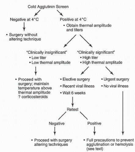
In summary, all patients undergoing hypothermic CPB should be screened preoperatively for cold agglutinins (19,20,21,23,28). If an initial screen is positive, the cold agglutinins should be characterized as to thermal amplitude and titer. Clinical symptoms should also be sought. A patient with low titer, low thermal amplitude, and clinically asymptomatic can tolerate CPB with moderate hypothermia at very low risk with little or no alteration in technique. For clinically significant cold agglutinins with high thermal amplitude and/or titer, if due to a transient viral illness, elective surgery may be postponed for several weeks in the hope that this will decrease cold agglutinins to insignificant levels. However, in severe cases in which the high-titer, high-thermal-amplitude cold agglutinins are not transient, precautions should be taken as outlined above to prevent an agglutination or hemolytic crisis (Fig. 24.2).


Hemoglobinopathy and Erythrocyte Disorders


Sickle Cell Trait and Disease

In the normal adult, hemoglobin A comprises 96% to 97% of all hemoglobin. Hemoglobin A is a tetramer composed of two α and two β-globin chains each associated with one oxygen carrying heme moiety. Sickle cell hemoglobinopathy (hemoglobin S) is a single-gene recessive abnormality in which the amino acid valine is substituted for the normally occurring glutamic acid at position 6 in the beta chain. It is thought that this single nucleotide polymorphism arose in central Africa primarily because the heterozygous form (sickle cell trait) confers some resistance to malaria, which is particularly endemic in that region. Sickle cell disease is the homozygous form of this hemoglobinopathy. There are a variety of subtypes of the condition. The percentage of hemoglobin S and hemoglobin A
varies with the individual, but sickle cell disease patients typically have predominantly hemoglobin S. This homozygous state is found in 0.15% of African-Americans and is associated with severe hemolytic anemia and vaso-occlusive phenomena, resulting from the increased blood viscosity that occurs when red blood cells aggregate and individually typically assume a sickle shape (29). Sickled cells have a very limited capacity to load and unload oxygen.


FIGURE 24.2. Algorithm for management of cold agglutinins in cardiopulmonary bypass.

In contrast, patients with the sickle cell trait have a lower percentage of hemoglobin S, accounting for 20% to 45% of their total hemoglobin. Approximately 8% of African-Americans carry the heterozygous recessive trait. These individuals have few clinical problems, and except for severe or provoked conditions, they rarely experience sickle cell crises. Red blood cell sickling results from deoxyhemoglobin formation. The tendency toward sickling increases with hypoxemia, acidosis, increased concentrations of 2,3-diphosphoglyceric acid, infection, hypothermia, and capillary stagnation. A hypertonic environment that may lead to crenation of normal red blood cells also will lead to sickling. Hemoglobin S demonstrates increased osmotic and mechanical fragility, making hemolysis more likely. A hypotonic environment will lyse red blood cells with increased osmotic fragility.

In patients with sickle cell disease, some sickling begins to appear at 85% hemoglobin oxygen saturation, and sickling of red blood cells is complete at 38% hemoglobin oxygen saturation. In patients with sickle cell trait, sickling begins at hemoglobin oxygen saturations of approximately 40%. Sickling is reversible to a degree, but if it is repeated, the sickled cells become permanently damaged, resulting in markedly increased fragility and a shortened cell lifespan. In addition to increased blood viscosity potentially causing vascular occlusion, sickling can cause endothelial cell injury in the microvasculature. This may activate the intrinsic clotting system and exacerbate the vaso-occlusive phenomenon (29,30,31,32).

The operative strategy in sickle cell patients is to prevent sickling and thereby prevent hemolysis or vaso-occlusive phenomena intraoperatively and postoperatively. Because sickling results from decreased hemoglobin oxygen saturation, maintaining adequate arterial oxygen tension assumes paramount importance. Adequate capillary perfusion with short capillary transit times and avoidance of low output states (to prevent low mixed venous hemoglobin oxygen saturations) are also important (33,34,35). Continuous measurement of arterial and mixed venous hemoglobin oxygen saturations help to maintain adequate oxygen saturations. Because sickle crises are frequent when high concentrations of hemoglobin S are present (i.e., homozygous patients), marked reduction of sickling can be achieved by relative dilution of hemoglobin S with respect to hemoglobin A. This can be accomplished by using preoperative or intraoperative exchange transfusions.

Preoperative exchange transfusions, particularly applicable in patients who are already anemic, not only increase the prevalence of hemoglobin A relative to hemoglobin S but also suppress the production of hemoglobin S. This therapy improves the oxygen-carrying capacity of the blood by correcting the anemia and deficiency of hemoglobin A. In nonanemic patients, exchange transfusion may be accomplished intraoperatively. This type of transfusion is usually performed by sequestering the initial CPB venous drainage from the patient after priming the extracorporeal circuit with whole blood containing hemoglobin A (29,35). The goal of exchange transfusion is to achieve a hemoglobin A fraction of 60% to 70%, which is also the level sought when treating a major sickle cell crisis (34).

Acidosis shifts the oxyhemoglobin dissociation curve to the right, which increases the tendency toward sickling. This holds true particularly in venous blood, where sickling is most often initiated. Arterial and mixed venous blood gases should be measured frequently and any developing acidosis aggressively treated with sodium bicarbonate (34,35). Hypoperfusion may result from hypothermia, administration of cardioplegia, diminished intravascular volume, poor patient positioning, tourniquets, low CPB systemic flows, or low cardiac output states. It is important to avoid hypoperfusion because of the tendency of blood to desaturate during the capillary and venous phase of circulation, resulting in low hemoglobin oxygen saturation and red blood cell sickling. Hypoperfusion can usually be prevented by maintaining adequate systemic CPB
flows and avoiding low cardiac output states both before and after bypass.

Localized sickling may occur in the heart during aortic cross-clamping because of the absence of coronary blood flow. This phenomenon may be avoided by flushing hemoglobin S out of the coronary arteries using either crystalloid cardioplegia or blood cardioplegia with a high fraction of hemoglobin A. Because mechanical prosthetic valves may predispose the patient to increased hemolysis, such valves are not recommended in these patients (35). Other means of avoiding mechanical blood trauma include minimizing the use of cardiotomy suction and venting. In patients with sickle cell disease, it appears advisable to minimize or avoid hypothermia during CPB. Despite the risks involved, numerous patients with homozygous sickle cell disease have successfully undergone CPB using the techniques described above (36,37,38,39,40,41,42,43,44,45). Successful cases have been described even using deep hypothermia with circulatory arrest (46).


Hereditary Spherocytosis

Hereditary spherocytosis, an autosomal dominant defect in red blood cell membranes, results in spherically shaped red blood cells that have increased osmotic and mechanical fragility. The usual treatment for hereditary spherocytosis resulting in hemolysis is splenectomy, which corrects the hemolysis and increases the shortened lifespan of the red blood cells to normal, although the cells retain their abnormal properties. Information describing CPB in these patients is limited. One case report describes a nonsplenectomized patient undergoing bypass with no apparent increase in blood destruction or in osmotic fragility over the baseline level (47). In addition, in a patient who had previously undergone splenectomy, no increase in hemolysis was noted during or after CPB, despite triple valve replacement using Bjork-Shiley mechanical valves (48). Other patients have had porcine valves inserted to minimize mechanically induced hemolysis. One study reported uneventful closure of an atrial septal defect in a 31-year-old with hereditary spherocytosis and suggested that a short CPB time was important in avoiding complications (49). Hereditary elliptocytosis is a condition thought to be similar to hereditary spherocytosis; there is infrequent hemolysis and anemia, and no specific precautions are recommended in these patients (50).


Thalassemia and G-6PD Deficiency

Thalassemia minor patients exhibit no increase in red blood cell fragility, and therefore one would expect no hemolysis. Anemia in these patients is treated with transfusions. Hemolysis has been induced by a number of drugs in patients with glucose-6-phosphate dehydrogenase deficiency (50). This gender-linked inherited deficiency is present in 10% to 15% of African-American males. Susceptible individuals may develop explosive hemolysis when they receive drugs such as antimalarials, quinidine, phenacetin, or sulfonamides. These drugs should be avoided in susceptible patients undergoing surgery, including those undergoing CPB.


Methemoglobin

Acute methemoglobinemia may result from increased production of methemoglobin to levels far exceeding the usual amount, which is less than 1% of total circulating hemoglobin (51). Secondary or acquired methemoglobinemia is almost always caused by poisoning with chemicals or drugs classified either as direct oxidants such as nitrites or indirect oxidants such as benzocaine. Other such medications include highdose methylene blue (52), nitroglycerin (53,54,55,56), nitroprusside, prilocaine, silver nitrate, sodium nitrate, flutamide (56), and sulfonamides.

The diagnosis of methemoglobinemia is made when cyanosis or oxygen desaturation occurs in the presence of an adequate arterial oxygen tension and is supported by a chocolate-brown color of blood rather than the usual dark blue of cyanosis (57). The diagnosis can be confirmed by spectrophotometry (58).

Treatment at times may need to precede definitive diagnosis. First, all probable offending drugs should be withdrawn. Next, maximal oxygen concentrations should be delivered to the oxygenator. If cyanosis or oxygen desaturation persists in the presence of high oxygen tension, pharmacologic treatment should begin. The drug of choice is methylene blue, 1 to 3 mg/kg administered in a 1% solution, which converts methemoglobin to active hemoglobin. The response to methylene blue is usually immediate and excellent, and because the treatment is relatively innocuous, its use should not be delayed. However, methylene blue may cause methemoglobinemia when a dose greater than 7 mg/kg is administered (59). If the patient fails to respond to methylene blue, the next line of treatment consists of high-dose vitamin C and, if necessary, exchange transfusion (53).


Polycythemia

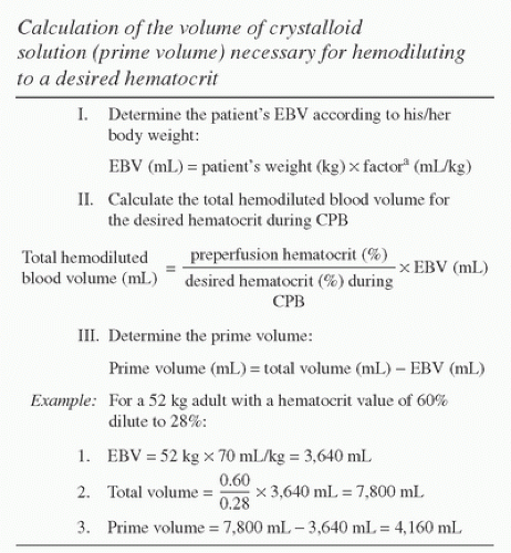
Polycythemia is defined as increased red blood cell mass. It occurs with cyanotic congenital cardiac defects as compensation for reduced oxygen delivery to tissues. The preoperative hematocrit in severe cases may exceed 70%, at which level blood viscosity is sufficiently high to compromise blood flow. Hemostatic abnormalities consistent with consumptive coagulopathy can occur (increased prothrombin and activated partial thromboplastin times, increased fibrin degradation products, thrombocytopenia, factor deficiencies, etc.) (60,61,62). Hemodilution beyond that normally used to prime the CPB circuit may better preserve the patient’s coagulation status, so this may be an ideal situation for withdrawal of autologous blood before CPB. The degree of hemodilution needed to achieve a desired patient/pump hematocrit can be calculated using a formula shown in Figure 24.3 (60). Polycythemia vera is a hematologic disease that carries an increased risk of myocardial infarction.


Religious Objections to Blood Transfusion

Avoidance of homologous blood transfusion is a desirable goal whenever CPB is used; with patients of the Jehovah’s Witness
faith it is mandatory because of their strict interpretation of the bible (63). More than 50 years ago, Cooley et al. (64) first reported the feasibility of open-heart surgery in this patient population. In 1977, Ott and Cooley (65) reported additional results in a large series of Jehovah’s Witness patients in whom no blood was transfused. However, there was a 10.7% mortality in those undergoing CPB (n = 39), with preoperative or postoperative anemia a contributing factor in 12 deaths.


FIGURE 24.3. Calculation of volume of crystalloid solution (cardiopulmonary bypass prime volume) necessary for hemodiluting to a desired hematocrit. EBV, estimated blood volume; CPB, cardiopulmonary bypass. aFactor (EBV/kg) is assumed to be 80 mL/kg for <10 kg body weight; 75 mL/kg for 10 to 20 kg of body weight; 70 mL/kg for >20 kg of body weight. The example addresses the polycythemic adult, but this condition is more prevalent in cyanotic pediatric patients with smaller required CPB circuit prime volumes. (From Milam JD, Austin SF, Nihill MR, et al. Use of sufficient hemodilution to prevent coagulopathies following surgical correction of cyanotic heart disease. J Thorac Cardiovasc Surg 1985;89:623-629, with permission.)

Using current low-prime membrane oxygenator circuits, intraoperative cell salvage, and reinfusion of shed mediastinal blood, CPB can be performed relatively safely, even in pediatric patients (63,66,67), reoperations, or those with complex anatomy (68). The lowest safe hematocrit on CPB is not known, but values of approximately 15% have been used successfully provided CPB systemic flows are maintained at levels to prevent development of metabolic acidosis.

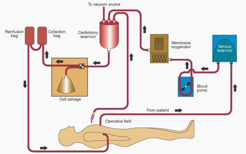
Most of these patients (but not all) who will accept the use of CPB will also accept the return of cell salvage products provided that continuity between removed blood and the patient’s vascular system has been maintained when cell salvage is used (69). Figure 24.4 shows how this can be accomplished pre- and post-bypass. We recommend having and documenting this specific discussion preoperatively. Some authors have further advocated use of heparin-bonded CPB circuits and lower levels of heparinization (activated clotting time [ACT] >280 seconds) with favorable results (70). Use of erythropoietin preoperatively to promote red blood cell production and antifibrinolytics perioperatively also have been advocated (71,72). Van Son et al. (67) enumerated management strategies to minimize blood loss in these patients (Table 24.2). Lee and Martin (73) wrote an excellent review of CPB management in this patient population.


Reoperative Surgery

Patients undergoing repeat cardiac surgery often present problems because of adhesions that make a second sternotomy and vascular access for CPB cannulation technically difficult. There is also an increased risk of encountering major bleeding during dissection because cardiac structures or vessels may be adherent to the chest wall, particularly if the pericardium has not been closed during the initial operation (74). Normal anatomic landmarks are often obliterated, prolonging adequate surgical exposure and CPB times. Because of adhesions necessitating sharp dissection, these patients tend to bleed more and have higher transfusion rates than first time cases (75). The surgeon should dissect as little as possible during reoperative surgery to minimize large disrupted tissue surface areas that will bleed. Use of an argon beam coagulator in these cases may also minimize bleeding problems associated with extensive dissection.

In some cases, groin cannulation of the femoral or iliac artery and femoral vein must be used to establish CPB. Placing an adequately sized femoral arterial cannula can usually be accomplished easily, but placement of an adequately sized venous cannula may be more difficult. New long, thin-walled, kink-resistant femoral venous cannulas are commercially available to permit positioning the cannula tip near the cavoatrial junction (76). However, because of their length, standard gravity siphonage may be inadequate to permit full CPB systemic flow. If maximizing the height differential between the patient’s heart and the CPB venous reservoir or repositioning the cannula does not improve venous drainage, flow may be augmented with a centrifugal pump placed in the venous line (77,78). Activation of the centrifugal pump will exert additional negative pressure beyond that obtainable by height differential alone with a concomitant modest increase in venous line flow. Alternatively, a hard-shell venous reservoir may have regulated vacuum applied to its interior to effect additional venous line flow using the same principles (79).

Both methods of augmented venous drainage require careful monitoring of venous line pressure so that excessive levels of vacuum are not created (80). Excessive vacuum can collapse vascular walls into the venous cannula openings, thus impeding or stopping venous line flow (81). Excessive
vacuum may be manifested by intermittent or staccato flow; in severe situations, the venous line may rhythmically jerk and relax as flow stops and is then reestablished with systemic venous return in the patient’s cavae or right atrium. Levels of hemolysis will also quickly rise if excessive vacuum is exerted in the venous line (82).


FIGURE 24.4. Schematic drawing of cardiopulmonary bypass (CPB) circuit for collection, processing, and reinfusion of blood after bypass while maintaining continuity with the patient’s circulation. The reinfusion bag (top left) should initially be back-filled with patient’s blood from an intravenous site to establish continuity with cell salvaged blood (from collection bag) before CPB. Cardiotomy suction can be used after bypass until protamine is administered with collected blood processed in the cell salvage system. Residual perfusate in the CPB circuit should be transferred to the cardiotomy reservoir and also processed by the cell-salvage system to minimize blood loss. A second cardiotomy reservoir (not shown) is used during CPB for conventional collection of suctioned and vent blood, which is drained into the venous reservoir. (Modified from Milan TP Jr, Whitmore J, Maddi R. Reoperative cardiac surgery in a Jehovah’s Witness: role of continuous cell salvage and in-line reinfusion. J Cardiothorac Anesth 1989;3:211-214, with permission.)

Establishing CPB via the groin vessels will afford the surgeon more control if massive bleeding in the chest is encountered. Alternatively, if the arterial cannula has been placed, CPB may be established using the pump suckers and a vent as a source of venous return (so-called sucker bypass). This will allow surgical control of bleeding and provide adequate decompression of the heart and preserve patient hemodynamics until the surgeon can place a conventional venous cannula in the right atrium or right atrium/inferior vena cava. The perfusionist should be prepared with additional cannulas, connectors, and tubing if CPB must be established emergently during reoperations (83). It must be recognized that the risk of aortic dissection is much greater when femoral arterial cannulation is used, which is discussed later in this chapter.

In summary, the number of patients presenting for reoperation is increasing. Increased surgical experience, use of antifibrinolytic drugs, and newer CPB technology can reduce the risk of morbidity and mortality associated with these procedures to levels approaching primary cardiac operation (84,85). Augmented venous drainage techniques have also been applied during minimally invasive cardiac surgery (86) and are discussed in greater detail subsequently.


Cardiopulmonary Bypass after Pneumonectomy

CPB techniques for patients who have undergone prior pneumonectomy have been addressed in the literature (87,88). The technical aspects of conducting CPB in such patients are not significantly different from those in patients who have had lobectomy or no pulmonary resection.

Hemodilution for CPB has been associated with a decrease in postoperative pulmonary problems theoretically from dilution of noxious blood elements or from avoidance of noxious elements (e.g., microaggregates present in homologous
blood). However, excessive hemodilution may predispose to pulmonary edema, which might be addressed by limiting hemodilution to a hematocrit fraction of more than 20% in postpneumonectomy patients.


TABLE 24.2. Perioperative measures to minimize blood loss in the Jehovah’s Witness patient


1.


Pretreat patient with synthetic erythropoietin if hematocrit <38%.


2.


Avoid cardiac catheterization, if possible.


3.


Administer antifibrinolytics perioperatively.


4.


Use low-energy electrocautery in chest wall, pericardial, and great vessel dissections.


5.


Use cell salvage system.


6.


Minimize pre-CPB fluid administration.


7.


Reduce CPB circuit priming volume.


8.


Use moderate CPB systemic flow rates and gentle cardiotomy suction.


9.


Use hemoconcentration to remove excess plasma water.


10.


Avoid deep levels of hypothermia if possible.


11.


Delay heparin neutralization until all bleeding sites have been secured (this will allow continued use of cardiotomy suction).


12.


Gradually return entire volume of residual perfusate to patient (after processing by cell salvage system).


13.


Administer postoperative iron supplements in patients with depleted red blood cell mass.


CPB, Cardiopulmonary bypass.


Derived from Von Son Jam, Hovaguimian H, Rao IM, et al. Strategies for repair of congenital heart defects in infants without the use of blood. Ann Thorac Surg 1995;59:384-388.


Blood transfusions should be avoided not only because of potential infections or transfusion reactions but also because of possible pulmonary damage from such elements as platelets and white blood cells. Washed packed red blood cells appear more appropriate when transfusion is indicated; return of shed blood should be avoided. When platelet transfusion is indicated, steroids and diphenhydramine should be used as pretreatment and leukocyte-depleted platelet concentrate should be used. Leukocyte reduction of all transfused residual perfusate will lessen the potential for pulmonary injury (89).

Technically, the position of the heart may be distorted because of contracted fibrothorax and/or hyperinflation of the remaining lung. This may lead to technical difficulty in gaining exposure, especially after left pneumonectomy. Remote cannulation from the femoral vessels has been helpful in some patients. Monitoring of pulmonary artery or left atrial pressure, with possible left ventricular venting, is important because strict control of the level of pulmonary capillary hydrostatic pressure is critical both intraoperatively and postoperatively to prevent pulmonary edema. Air emboli in the pulmonary circuit would seemingly be less well tolerated, and this is addressed by standard de-airing techniques. Time on CPB directly correlates with postoperative lung water; therefore, at times, a less complete coronary revascularization may be preferable to incurring a long CPB time. For coronary bypass surgery, the proximal anastomoses may be performed off pump or with low-flow partial CPB.


Minimally Invasive Surgery

A full discussion of the use of CPB in support of all minimally invasive cardiac surgery, such as port access and robot-assisted procedures, is beyond the intended scope of this chapter. More detailed discussion of these techniques may be found in Chapter 7. However, two features commonly used with CPB support for these minimally invasive approaches, namely cannulation of femoral vessels and management of venous drainage warrant specific consideration here.

Femoral arterial cannulation is recognized to have a somewhat different risk profile than ascending aortic cannulation. Specifically there may be a higher occurrence of aortic dissection and other complications associated with the more traditional blind insertion of a relatively short femoral artery cannula. Although published literature is sparse a more reasonable approach is the use of a modified Seldinger technique with a guidewire and sequential dilation of the vessel to the point where it will accept the appropriate cannula size capable of delivering expected required flow rates within acceptable circuit pressure ranges. Visualization of the initial guidewire in the distal thoracic aorta using intraoperative TEE is helpful in assuring that the cannula will be located within the aortic lumen.

Femoral vein cannulation typically utilizes a long cannula that is passed to the point of the superior vena cava/right atrial junction. This position may be approximated by surface measurements but correct positioning is greatly assisted by direct visualization of first the guide wire and subsequently the venous cannula using intraoperative TEE. Because of the length and relatively small diameter of these cannulas, the conventional siphon pressure gradient may not be sufficient to generate adequate venous drainage blood flow. If this occurs, then venous flow may be augmented significantly by application of vacuum to the (hard shell) venous reservoir. Use of the minimum vacuum pressure necessary to produce the necessary increment in venous drainage is recommended. Higher venous vacuum can produce air entrainment if vascular openings to the ambient environment are present. Hemodynamic monitoring catheters, unrecognized intracardiac defects, cannula misplacement, and other iatrogenic causes have been implicated. Air entrainment can be sufficient to worsen (or totally obstruct) venous drainage, or in a worst-case scenario air can pass from the venous circuitry into the systemic perfusion side of the circuit with potentially disastrous results. Further discussion of air embolization may be found in a subsequent section of this chapter. Despite the apparent logic of further increasing the venous vacuum to increase venous drainage when air entrainment occurs to accelerate passage
of the air and improve flow, the logic is flawed. Greater negative pressure will only serve to increase air entrainment and worsen the issues. The preferred approach would be to identify the source of the air entrainment and prevent further entrainment.

Jun 7, 2016 | Posted by in RESPIRATORY | Comments Off on Management of Unusual Problems Encountered during Procedures that Require the Use of Cardiopulmonary Bypass

Full access? Get Clinical Tree

Get Clinical Tree app for offline access