Management of Heart Failure

23 Management of Heart Failure


Heart failure (HF) is a complex clinical syndrome that can result from any structural or functional cardiac disorder that impairs the ability of the ventricle to fill with (diastolic HF) or eject (systolic HF) blood. Most commonly, HF results from myocardial muscle dysfunction with accompanying dilation or hypertrophy of the left ventricle (LV), remodeling, and neurohormonal activation.


There are an estimated 23 million people with HF worldwide. In the United States, the prevalence is high. An estimated 5.3 million Americans have HF, and 660,000 new cases of HF are diagnosed per year. The incidence of HF increases significantly with age. HF results in over 1 million hospitalizations annually and is the most common cause of hospitalization for patients aged 65 years and older. The annual health care cost of patients with HF is projected to exceed $34 billion in 2008. With appropriate therapy, patients with HF can be stabilized and have significant improvement in their symptoms. However, despite therapeutic advances, the mortality rate is high, about 50% at 5 years. HF is recorded in one out of every eight death certificates. It is likely that the broader use of evidence-based approaches for the treatment of patients with HF will lead to reduction in mortality. More aggressive efforts for risk factor modification, especially for coronary heart disease risk factors, are of importance given that HF following myocardial infarction (MI) is common. Studies have demonstrated that treating hypertension, vascular disease, or high-risk diabetics significantly reduces the incidence and development of HF.


Risk factors for developing HF include a history of atherosclerotic vascular disease, smoking, hypertension, diabetes, obesity, valvular disease, hyperlipidemia, physical inactivity, excessive alcohol intake, exposure to cardiotoxins, family history of cardiomyopathy, and sleep-disordered breathing.


The American College of Cardiology/American Heart Association update in 2005 presented a new combined clinical and pathophysiologic classification for HF based on four stages:


The focus of this chapter is on those individuals who do have evidence of myocardial dysfunction or HF—patients in stages B, C, and D.


Etiology and Pathogenesis


Coronary artery disease (CAD) accounts for 50% of the incidence of HF worldwide. Patients with a previous MI can develop both decreased systolic performance and diastolic impairment due to interstitial fibrosis and scar formation. Hibernating myocardium due to severe CAD can also cause systolic HF, which is potentially reversible with revascularization. Hypertension is a common cause of HF, especially in African Americans and older women. Valvular heart disease accounts for approximately 10% to 12% of cases of HF. A common cause of initially unexplained HF (following exclusion of CAD) is idiopathic cardiomyopathy. Familial cardiomyopathies may account for up to one third of cardiomyopathies thought to be idiopathic. Other etiologies of dilated cardiomyopathy (Chapter 18) include thyroid disease, chemotherapy (anthracyclines, e.g., doxorubicin and trastuzumab [Herceptin]), myocarditis (Chapter 22), infection due to HIV, diabetes, alcohol, cocaine, connective tissue disease, systemic lupus erythematosus, peripartum cardiomyopathy, and arrhythmias. Hypertrophic (Chapter 19) and restrictive (Chapter 20) cardiomyopathies can cause HF, but this is less common.


Systolic Heart Failure


Systolic HF (ejection fraction [EF] = 40%) results in a reduction in cardiac output that is perceived as “hypovolemia” by the kidneys and triggers activation of the renin-angiotensin-aldosterone system (RAAS). With RAAS activation, salt and water retention occurs. Initially, this results in increased preload, transiently improving cardiac output. Over longer periods of time, chronic activation of the RAAS results in volume overload and symptoms of HF. Declining blood pressure due to decreased cardiac output also triggers activation of the sympathetic nervous system. Increased levels of angiotensin II, aldosterone, catecholamines, endothelin, and vasopressin result in systemic vasoconstriction. The short-term benefit of vasoconstriction—increased perfusion of critical organs—is followed by worsening HF due to chronically increased LV afterload. Sympathetic nervous system activation can also precipitate ventricular arrhythmias, a common cause of death in patients with HF.


HF generally follows an injury to the myocardium (due to ischemia, a toxic effect, or an increased volume or pressure load on the LV). LV remodeling, a maladaptive response, follows, with resulting changes in cardiac size, shape, and function (Fig. 23-1). Myocyte length increases, with a resulting increase in chamber volume, which preserves stroke volume. Myocyte hypertrophy can also occur, along with a loss of myocytes due to apoptosis or necrosis, and fibroblast proliferation and fibrosis. The heart remodels eccentrically in systolic HF, becoming less elliptical and more spherical and dilated. The mitral valve annulus often becomes dilated, resulting in mitral regurgitation and further increased wall stress.
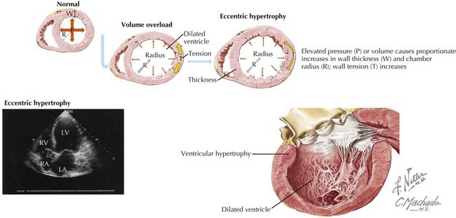


The success of angiotensin-converting enzyme inhibitors (ACE-Is), angiotensin II receptor blockers (ARBs), β-blockers, and aldosterone antagonists in reducing mortality in patients with HF is in large part due to their ability to block neurohormonal activation and subsequently attenuate and even reverse remodeling.


Diastolic Heart Failure


Diastolic heart failure (DHF) is characterized by normal LV volume, concentric remodeling, normal LV systolic function, and abnormalities of diastolic function. DHF accounts for 40% to 50% or more of HF cases. DHF affects older patients, especially women. Ischemic heart disease and hypertension are the most common causes of isolated DHF. In the typical patient with DHF, the ventricular size is normal. However, if DHF occurs as a result of mitral or aortic valve regurgitation, or because of a high-output state (such as anemia or thiamine deficiency), ventricular dilation may be present. The morbidity and mortality of patients with DHF is similar to that of patients with HF due to systolic dysfunction.


Hypertrophic and restrictive cardiomyopathies can result in a clinical presentation consistent with DHF (see Chapters 19 and 20), as can constrictive pericarditis. Indeed, distinguishing these entities can be difficult, requiring extensive noninvasive and invasive hemodynamic assessment.


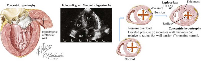
DHF is generally characterized by a normal end-diastolic volume, hypertrophy of the cardiomyocytes, and increased wall thickness resulting in a concentric pattern of LV remodeling as compared with the increased cardiomyocyte length, increased end-diastolic volume, and eccentric remodeling seen in systolic HF (Fig. 23-2). There is increased extracellular matrix, abnormal calcium handling, and activation of the RAAS and sympathetic nervous system. Together, these pathophysiologic changes result in impaired ventricular relaxation, high LV diastolic pressure, high left atrial filling pressures, and resulting symptoms and signs of HF.


Clinical Presentation


The presentation of patients with HF includes signs and symptoms of pulmonary congestion, systemic fluid retention, exercise intolerance, or inadequate organ perfusion. Symptoms include dyspnea on exertion, exercise intolerance, orthopnea, paroxysmal nocturnal dyspnea, cough, chest pain that may or may not represent angina, weakness, fatigue, volume overload or pulmonary hypertension, nocturia, insomnia, depression, and weight gain. Patients with end-stage disease may also complain of nausea, abdominal pain, oliguria, confusion, and weight loss. Physical examination findings that should be assessed include jugular venous pressure, rales, wheezing, pleural effusion, displaced point of maximal intensity, right ventricular heave, increased intensity of P2 due to pulmonary hypertension, S3, S4, murmurs, hepatomegaly, hepatojugular reflux, low-volume pulses, and peripheral edema. Patients with end-stage disease may also exhibit pulsus alternans, ascites, cool, pale extremities, and cachexia.


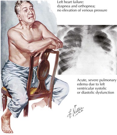
The clinical presentation may be indistinguishable between patients with systolic and diastolic HF (Fig. 23-3). The cardiac silhouette is usually enlarged in both circumstances, with cardiomegaly due to ventricular dilation in systolic HF and from hypertrophy in patients with DHF. An assessment of LV function is essential for determining the appropriate approach to therapy.


Differential Diagnosis


The difficulty in arriving at a new diagnosis of HF lies in its vague symptoms and examination mimickers (Box 23-1). Symptoms of dyspnea and exercise intolerance can be attributed to many diagnoses: lung disease (including chronic obstructive lung disease, reactive airways diseases, thromboembolic pulmonary disease, and pulmonary hypertension), thyroid disease, arrhythmias, anemia, obesity, deconditioning, and cognitive disorders. Signs of volume overload are not specific to HF. Sodium-avid states of nephrosis and cirrhosis, as well as pericardial disease, can present with similar findings of jugular venous distention, hepatomegaly, and edema.


Diagnostic Approach


The diagnosis is made by taking a careful history, performing a directed examination, and assessing systolic and diastolic ventricular function. Laboratory evaluation (electrolytes, glucose, calcium, magnesium, lipid profile, complete blood count, albumin, liver functions tests, urinalysis, thyroid function), ECG, CXR, and pulmonary function testing will eliminate most noncardiac diagnoses.


Additional directed tests include iron studies (ferritin and total iron binding capacity) to screen for hereditary hemochromatosis, antinuclear antibody and other serologic tests for lupus, viral serologies and antimyosin antibody if myocarditis is suspected, evaluation for pheochromocytoma, serum protein electrophoresis, urine protein electrophoresis, and thiamine, carnitine, and selenium levels.


Measurement of serum brain natriuretic peptide (BNP >400 pg/mL) or N-terminal prohormone BNP (pro-BNP >450 pg/mL in individuals younger than 50 years, >900 pg/mL in individuals 50–75 years old, or >1800 pg/mL in patients over 75 years old) can be very helpful in the acute setting. These markers correlate with elevated filling pressures and are particularly helpful in the evaluation of patients with dyspnea. Although an elevated BNP or pro-BNP level does not rule out pulmonary causes of dyspnea, normal levels (BNP <100 pg/mL or proBNP <300 pg/mL) argue against HF as the predominant cause of dyspnea. Even though levels are generally higher in cases of systolic HF, these tests cannot distinguish between systolic and DHF.


Determining the Type and Degree of Left Ventricle Dysfunction


Echocardiography is the most common method for initial assessment of LV function. EF, valve function, hypertrophy, and diastolic function can all be assessed. Most patients with DHF have impaired LV relaxation, with or without a quantifiable reduction in LV compliance, and preserved EF. The most reproducible and validated method of diagnosing diastolic dysfunction combines echocardiographic two-dimensional M-mode Doppler measurements of mitral valve inflow with the sensitive, relatively load-independent measure of LV relaxation (e′ velocity) obtained by tissue Doppler imaging of the mitral annulus. This approach has resulted in four classifications of diastolic function: normal, mild dysfunction (impaired relaxation, normal filling pressure), moderate dysfunction (impaired relaxation or pseudonormal with moderately elevated filling pressure), and severe dysfunction (restrictive) (Fig. 23-4). Reversibility can be determined with the Valsalva maneuver. An E/e′ ratio that exceeds 15 correlates with elevated filling pressure.
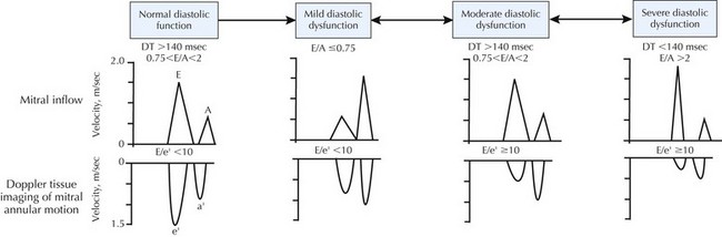


Radionuclide ventriculography can be used to determine EF in obese patients and in those with significant chronic obstructive pulmonary disease. The first-pass technique can quantify right ventricular EF as well. Cardiac MRI is a newer imaging modality that allows very accurate assessment of LV function (and EF) in all patients, provides an assessment of myocardial viability, and can identify infiltrative disease. In recent years it has become clear that there is a risk in gadolinium administration to patients with moderate to severe kidney disease, which is common in HF, including patients on dialysis. In these patients, gadolinium administration is associated with the severe syndrome of nephrogenic systemic fibrosis. Therefore, gadolinium administration should be avoided in these patients. Pacemakers and defibrillators are contraindications for MRI.


Jun 12, 2016 | Posted by in CARDIOLOGY | Comments Off on Management of Heart Failure

Full access? Get Clinical Tree

Get Clinical Tree app for offline access