16 Malignant Pleural Effusions Secondary to Metastatic Neoplasms
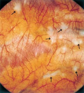
Fig. 16.1
Malignant pleural effusion from metastatic uterine carcinoma. After drainage of 800 mL of serous effusion, numerous, whitish nodules on both visceral and parietal pleura. The photograph shows a section of the chest wall with marked hyperemia. The horizontally oriented ribs and intercostal spaces are noted as well as numerous pale tumor nodules (→) which in part infiltrate the surrounding pleura in a radial fashion.
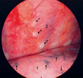
Fig. 16.2
Malignant pleural effusion from metastatic cancer of unknown origin. After drainage of 1800 mL of serous effusion, normal surface of the right lower lobe (1) with anthracotic pigmentation (→). Whitish small tumor nodules ( ) on the posterior chest wall (2).
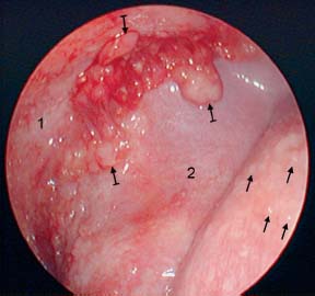
Fig. 16.3
Malignant pleural effusion from metastatic breast cancer. After drainage of 900 mL of serous effusion: small tumor nodules on the surface of the left lower lobe (→) and slightly larger nodules ( ) on the posterior chest wall (1) close to the diaphragm (2).
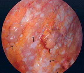
Fig. 16.4
Bilateral pleural effusions from metastatic breast cancer. After drainage of 1700 mL of serous effusion: nodular (→) and flat, whitish ( ) tumor lesions on the posterior chest wall (and on the diaphragm).
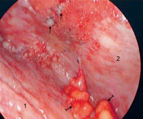
Fig. 16.5
Malignant pleural effusion from metastatic breast cancer. After drainage of 2800 mL of serous effusion: small and middle-sized tumor nodules on the diaphragm (1) and the chest wall (2), in part anthracotically pigmented (→). Fatty tissue with tumor infiltration ( ).
).
Stay updated, free articles. Join our Telegram channel

Full access? Get Clinical Tree


