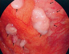
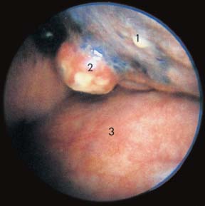
14 Malignant Pleural Effusions due to Lung Cancer Fig. 14.1 a Malignant pleural effusion due to small-cell lung cancer. After drainage of 4200 mL of serous effusion, the view toward the apex of the right thoracic cavity. Upper lobe (1) completely infiltrated by tumor. Lower lobe (2) partially infiltrated (→). Parietal pleura with the first three ribs and the intercostal spaces appear normal. b Malignant pleural effusion due to small-cell lung cancer. Same patient as in a: several small to middle-sized tumor nodules (→) on the anterior chest wall and in the fatty tissue . Fig. 14.2 Malignant pleural effusion due to small-cell lung cancer (recurrent under second-line chemotherapy). After drainage of 2300 mL of serous effusion, there is ubiquitous tumor growth on the right visceral and parietal pleura; here middle-sized nodules (→) and flat infiltrations () on the posterior chest-wall pleura. Fig. 14.3 Malignant pleural effusion due to small-cell lung cancer invading into the pleural space. After drainage of 500 mL of clear serous effusion, the base of the right lower lobe (1) is retracted cranially. One sees the hazelnut-sized, yellowish necrotic tumor nodule breaking through into the pleural cavity (2). The diaphragm (3) shows numerous dilated vessels and very discreet, barely visible, small nodules. Only gold members can continue reading. Log In or Register to continue Share this:Click to share on Twitter (Opens in new window)Click to share on Facebook (Opens in new window) Related Related posts: Medical Thoracoscopy/Pleuroscopy in Children Teaching Methods Tuberculous Pleural Effusions Pleural Effusions due to Diffuse Malignant Mesothelioma and Asbestos-related Pleural Diseases Stay updated, free articles. Join our Telegram channel Join Tags: Medical Thoracoscopy Pleuroscopy Manual and Atlas Mar 12, 2017 | Posted by admin in RESPIRATORY | Comments Off on Malignant Pleural Effusions due to Lung Cancer Full access? Get Clinical Tree
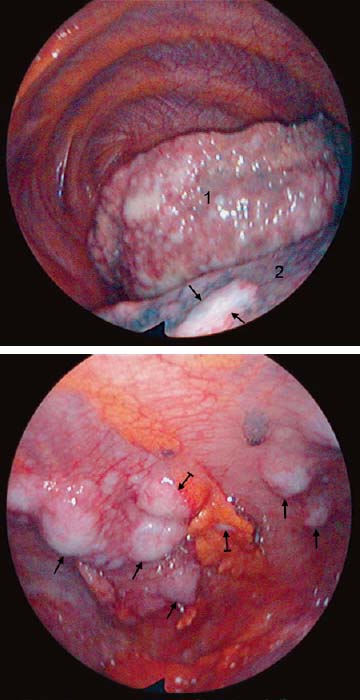
14 Malignant Pleural Effusions due to Lung Cancer Fig. 14.1 a Malignant pleural effusion due to small-cell lung cancer. After drainage of 4200 mL of serous effusion, the view toward the apex of the right thoracic cavity. Upper lobe (1) completely infiltrated by tumor. Lower lobe (2) partially infiltrated (→). Parietal pleura with the first three ribs and the intercostal spaces appear normal. b Malignant pleural effusion due to small-cell lung cancer. Same patient as in a: several small to middle-sized tumor nodules (→) on the anterior chest wall and in the fatty tissue . Fig. 14.2 Malignant pleural effusion due to small-cell lung cancer (recurrent under second-line chemotherapy). After drainage of 2300 mL of serous effusion, there is ubiquitous tumor growth on the right visceral and parietal pleura; here middle-sized nodules (→) and flat infiltrations () on the posterior chest-wall pleura. Fig. 14.3 Malignant pleural effusion due to small-cell lung cancer invading into the pleural space. After drainage of 500 mL of clear serous effusion, the base of the right lower lobe (1) is retracted cranially. One sees the hazelnut-sized, yellowish necrotic tumor nodule breaking through into the pleural cavity (2). The diaphragm (3) shows numerous dilated vessels and very discreet, barely visible, small nodules. Only gold members can continue reading. Log In or Register to continue Share this:Click to share on Twitter (Opens in new window)Click to share on Facebook (Opens in new window) Related Related posts: Medical Thoracoscopy/Pleuroscopy in Children Teaching Methods Tuberculous Pleural Effusions Pleural Effusions due to Diffuse Malignant Mesothelioma and Asbestos-related Pleural Diseases Stay updated, free articles. Join our Telegram channel Join