Utilization of heparin-coated circuits
Utilization of synthetic co-polymer(methacrylate)-coated circuits
Utilization of phosphorylcholine-coated circuits
Use of ultrafiltration techniques
Use of leukocytes filters
Use of the Drew-Anderson techniquea
Lung perfusion with controlled perfusion pressure
Lung perfusion with antibiotics and antiinflammatory solutions
Lung perfusion with pulsatile flow
Lung perfusion with low-volume ventilation
Inhalation of NO
Inhalation of CO
Miniaturized cardiopulmonary circuit
Management of hemodilution
Monoclonal anticytokine antibodies
The role of two substances in lung protection – protein C and adenosine – has been extensively addressed in recent papers worldwide. Thus, understanding their mechanism of action and the effective impact on pulmonary function will allow heart surgeons to attenuate the inflammatory response and ischemia-reperfusion injury during heart surgery [10].
Protein C has an activated form – activated protein C; this is a natural anticoagulant generated by the thrombin-thrombomodulin complex on endothelial cells. In addition to the anticoagulant role, this activated protein has antiinflammatory properties as it downregulates proinflammatory cytokines and regulates cardiopulmonary bypass-induced neutrophil activation [10, 11].
Yamakazi et al. [12] designed intriguing experimental research depicting the inflammatory and functional benefits of using activated protein C in heart surgery requiring cardiopulmonary bypass. These authors defined three groups as follows: control, DIP (intravenous inactive derivative of activated protein C), and APC (intravenous activated protein C). Histological analysis, arterial oxygen pressure, proinflammatory marker concentration, and CD11b expression are shown in Figs. 33.1, 33.2, 33.3, and 33.4. These figures allow us to suggest that, in terms of pulmonary function, activated protein C can play a protective role, minimizing inflammatory injury and providing functional balance [12].
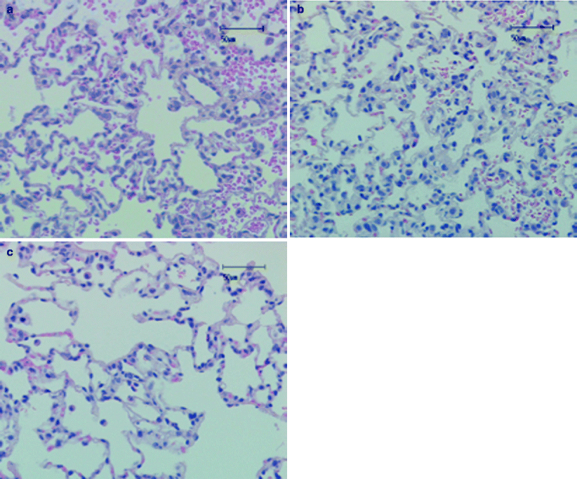
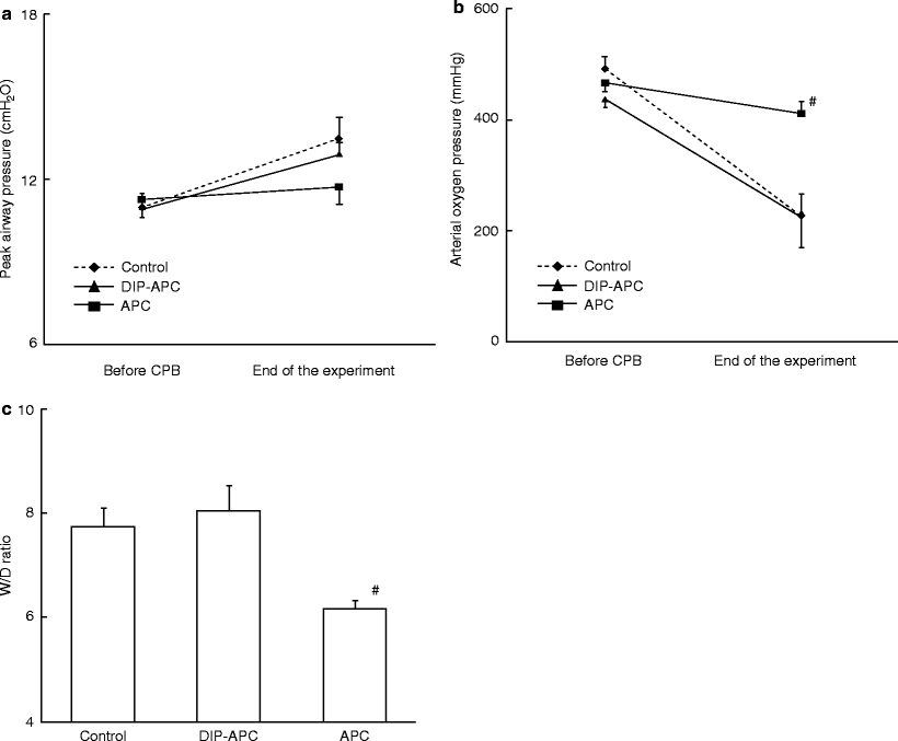
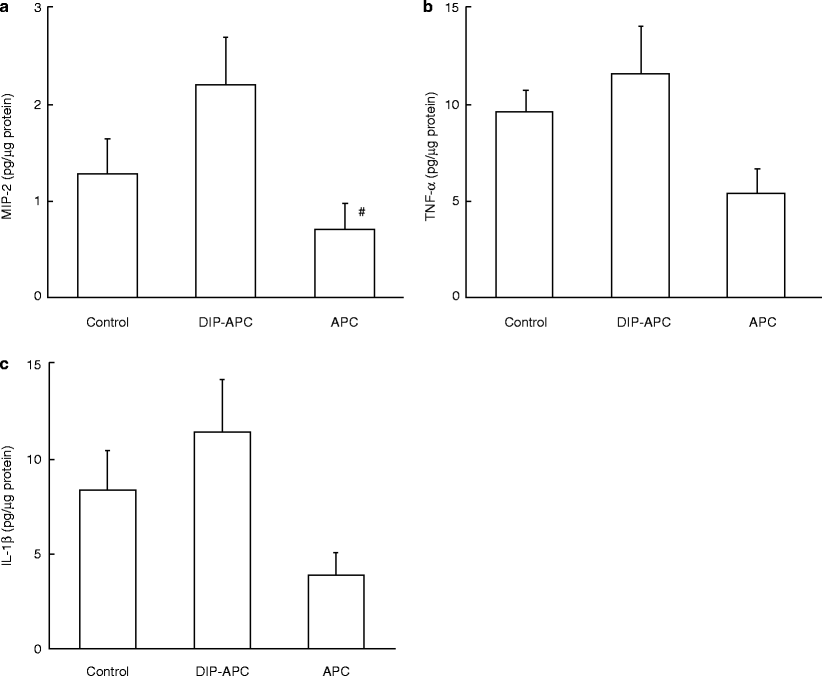


Fig. 33.1
Microphotograph of the left lung. The left lung section in each group was stained with hematoxylin & eosin. Lungs in the control and DIP groups show interstitial edema, alveolar hemorrhage, and severe neutrophil accumulation. Inflammatory change in the APC group lung is minimized. Original magnification: x200. Black bar = 50 mm. Control (a), DIP (b), and APC (c) groups
Fig. 33.2
(a) Peak airway pressure before CPB and at the end of the experiment. The APC group shows a relatively lower value than the other groups at the end of the experiment, although there are no significant differences among the groups. Results are expressed as mean ± standard error of the mean (SEM). (b) Arterial oxygen pressure before CPB and at the end of the experiment. The APC group shows a significantly higher value than the other groups at the end of the experiment. Results are expressed as mean ± SEM. #P < 0.01 versus the control or DIP group. (c) W/D weight ratio of the left lung. The APC group shows a significantly lower value than the other groups. Results are expressed as mean ± SEM. #P < 0.01 versus the control or DIP group. APC activated protein C, CPB cardiopulmonary bypass, DIP diisopropyl fluorophosphate, W/D wet to dry
Fig. 33.3
(a) Tissue MIP-2 concentration in the left lung. The APC group shows a relatively lower value than the control group and a significantly lower value than the DIP group. Results are expressed as mean ± SEM. #P < 0.01 versus the DIP group. (b) Tissue TNF-a concentration in the left lung. The APC group shows a relatively lower value than the other groups, although there are no significant differences among the groups. Results are expressed as mean ± SEM. (c) Tissue IL-1b concentration in the left lung. The APC group shows a relatively lower value than the other groups, although there are no significant differences among the groups. Results are expressed as mean ± SEM. DIP diisopropyl fluorophosphate, APC activated protein C, TNF tumor necrosis factor, MIP macrophage inflammatory protein, IL interleukin
< div class='tao-gold-member'>
Only gold members can continue reading. Log In or Register to continue
Stay updated, free articles. Join our Telegram channel
Full access? Get Clinical Tree
