Lung Cancer: Epidemiology, Surgical Pathology, and Molecular Biology

Chapter 65 Lung Cancer


Epidemiology, Surgical Pathology, and Molecular Biology


Epidemiology


Incidence and Survival


Lung cancer currently is the highest cause of cancer mortality in the United States and even surpasses the sum of the next four cancer types in both men and women. In terms of incidence, (the number of new cases of cancer in a given year), lung cancer is second only to breast cancer in women and prostate cancer in men. In the United States, estimates of new cases of lung cancer for 2010 were approximately 116,000 in men and 105,000 in women, with approximately 160,000 deaths. Lung cancer deaths accounted for 31% and 27% of overall cancer deaths in men and women, respectively, and are more numerous than deaths due to breast, prostate, and colon cancers combined. In terms of overall causes of death in men, lung cancer is the second most common cause of death, after vascular disease, with deaths due to heart attacks and stroke. The overall 5-year lung cancer survival rate is between 15% and 20% as a consequence of its late stage at onset of symptoms and relatively aggressive clinical behavior. Like other malignancies, lung cancer is a disease of aging, with increasing rates in persons older than 50 years of age, and rarely is diagnosed in those younger than 40 years. Graphing lung cancer incidence according to chronologic age of diagnosis on a logarithmic scale demonstrates a straight line for both men and women, indicating that the carcinogenic pathways probably are similar for both genders and that steroid hormones (estrogen and androgens) do not play a major role in carcinogenesis.


Lung cancer more typically is present in men than in women, and in African Americans than in white Americans; these disparities in incidence probably are due to smoking patterns. The disparity extends to younger persons as well; young African American men and women have significantly higher lung cancer rates than their white counterparts. The incidence of lung cancer in adult African American and in white American male adults in 2008 was 101 and 69 per 100,000, respectively. Both African American and white American women had similar cancer incidences of approximately 55 cases per 100,000. Other ethnic and racial groups, such as Hispanic persons, Native Americans, and Asian or South Pacific Americans have a lower incidence than that for white Americans.


Lung cancer incidence is highest in the developed nations of North America (United States and Canada), the European Union, and countries of the former Soviet Union, where cigarette availability and smoking rates are highest. In 2008, lung cancer was diagnosed worldwide in approximately 1.6 million people and led to cancer deaths in 1.4 million, and a majority of new cases are reported from China and less developed countries. Since the 1970s, lung cancer incidence has been decreasing in the United States, Canada, United Kingdom, and Australia but has been increasing in Japan and India. Unfortunately, during this same time, lung cancer incidence has been increasing among women in the United States, Canada, Denmark, Norway, and Sweden. Worldwide, lung cancer incidence is more than double for men relative to women, and the relative risk is even higher in Western Asia, Central and Southern America, and Southern Africa.


Although lung cancer is increasing in large developing nations, such as China and India, the socioeconomic demographic features are important in appreciating lung cancer incidence. The risk of lung cancer is inversely associated with education and income and appears to be more closely linked to smoking than risk of disease from occupational or environmental exposures. Data from developing nations are not as robust and accurate as those from Western countries, and analysis of the socioeconomic setting of smoking also involves confounding variables, such as local diet and environmental carcinogens and inhalant exposures.


When the mortality rates (deaths due to disease within a calendar year) for lung cancer in women and men are plotted against the calendar years for the last 50 years, one notes a marked difference in the pattern of the curves (Figure 65-1). The incidence rates for men show a marked rise from the early 1950s to the beginning of the 1990s, whereas rates for women lag by about a quarter of a century and then show an identical rise, essentially parallel to those for men, and continuing into the 21st century. By the 1950s, death due to lung cancer far exceeded prostate and colon cancer deaths among men, and by 1990, lung cancer deaths among women exceeded breast and gynecologic cancer deaths. Usually cancer mortality follows cancer incidence; however, in the case of lung cancer, owing to the very high cancer fatality rate (mortality from cancer for persons given a diagnosis of cancer), lung cancer mortality exceeds the most common cancers in both men and women. As a result of the Surgeon General’s 1964 report on smoking, men began to stop smoking in the succeeding 20 to 30 years; the incidence and mortality of lung cancer began to decline from a peak in the early 1990s to a level that approximates the mortality rate of the 1970s. The phenomenon of social smoking among women lagged behind that for men; subsequently, both the incidence of lung cancer in women and its associated mortality continue to rise into the present day.


image

Figure 65-1 A, SEER incidence and U.S. death rates for cancer of the lung and bronchus (men and women). Joinpoint Regression analyses for whites and blacks from 1975 to 2007 and for Asian/Pacific Islanders, American Indians/Alaska Natives, and Hispanics from 1992 to 2007. API, Asian Pacific Islander; AI/AN, American Indian/Alaska Native. B, SEER-delay adjusted rates for cancer of the lung and bronchus mortality differences among men and women of all races in the United States (1975-2007). SEER, Surveillance, Epidemiology, and End Results [Program].


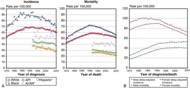
(A, Incidence data for whites and blacks are from the SEER nine areas—San Francisco, Connecticut, Detroit, Hawaii, Iowa, New Mexico, Seattle, Utah, Atlanta. Incidence data for Asian/Pacific Islanders, American Indians/Alaska Natives, and Hispanics are from the SEER thirteen areas—the SEER nine areas plus San Jose–Monterey, Los Angeles, Alaska/Native Registry, and Rural Georgia. Mortality data are from U.S. mortality files, National Center for Health Statistics, Centers for Disease Control and Prevention [CDC]. Rates are age-adjusted to the 2000 U.S. standard population [19 age groups, Census P25-1103]. Regression lines are calculated using the Joinpoint Regression program Version 3.4.3, April 2010, National Cancer Institute. Joinpoint analyses for whites and blacks during the 1975-2007 period allow a maximum of 4 joinpoints. Analyses for other ethnic groups during the period 1992 to 2007 allow a maximum of 2 joinpoints. *Rates for American Indians/Alaska Natives are based on the contract health service delivery area [CHSDA] counties. †Hispanic is not mutually exclusive of whites, blacks, Asians/Pacific Islanders, and American Indians/Alaska Natives. Incidence data for Hispanics are based on the NHIA [North American Association of Central Cancer Registries/NAACCR Hispanic Identification Algorithm] and exclude cases from the Alaska Native Registry. Mortality data for Hispanics exclude cases from Connecticut, the District of Columbia, Maine, Maryland, Minnesota, New Hampshire, New York, North Dakota, Oklahoma, and Vermont. B, Data from SEER nine areas and U.S. mortality files [National Center for Health Statistics, CDC]. Rates are age-adjusted to the 2000 U.S. standard population [19 age groups, Census P25-1103]. Regression lines are calculated using the Joinpoint Regression Program Version 3.4.3, April 2010, National Cancer Institute [http://seer.cancer.gov/statfacts/html/lungb.html].


Although lung cancer typically refers to the malignant bronchogenic epithelial tumors of the lung, namely, squamous cell carcinoma, adenocarcinoma, large cell carcinoma, and small cell (neuroendocrine) carcinoma, the relative incidence, distribution, and frequency of these tumors over time are characterized by marked differences. In the 1970s, squamous cell carcinoma predominated in incidence over the other bronchogenic carcinomas. Of interest, the age-adjusted incidence rate of adenocarcinoma, among both men and women, continued to rise, so that beginning in the late 1990s and continuing into the early 2000s, adenocarcinomas have become the most frequently encountered lung cancers. The two histologic subgroups of lung cancer most strongly associated with smoking, squamous cell carcinoma and small cell carcinoma, have shown a decrease in their overall incidence, reflecting the trend of decreased cigarette smoking among men. The relative percentages of the major lung cancer histologic types are adenocarcinoma 38%, squamous cell carcinoma 20%, small cell (neuroendocrine) carcinoma 13%, and large cell carcinoma 5%. The remainder are composed of variant carcinomas, sarcomatoid (spindle cell) carcinomas, salivary-type carcinomas, neuroendocrine-carcinoid carcinomas, and others.


The histologic sequence eventuating in squamous cell carcinoma begins with smoking-induced replacement of respiratory epithelium with bronchial squamous metaplasia. Additional cellular and molecular events lead to squamous dysplasia, in situ carcinoma, and ultimately, invasive squamous cell carcinoma. Small cell carcinoma does not have a clearly defined precursor lesion, although it is presumed to be the Kulchitsky cell; however, small cell carcinomas often arise in a setting of squamous metaplasia and dysplasia, indicative of the effects of the smoking environment. Adenocarcinoma, a histologic subtype characterized by formation of malignant-appearing glands, papillae, and intracytoplasmic mucin, also arises in the context of cigarette smoking, yet approximately 25% of lung cancer cases are not attributable to direct smoking. These tumors typically are located peripherally and may be associated with scars that in some cases may be preexisting but also may represent a host desmoplastic response to the tumor. As a histologic subtype, adenocarcinomas of the lung continue to increase in incidence among both men and women.


Etiology and Risk Factors


The major risk factor associated with lung cancer in a majority of cases is cigarette smoking. The probability that a diagnosis of lung cancer will be made in a nonsmoker is less than 15%. The case for cigarette smoking as a major cause of lung cancer is made by strong epidemiologic evidence and by significant results of animal studies. Both case-control and cohort studies have demonstrated the strong association of smoking with lung cancer and have shown that the association is dose-dependent and linked to current versus past smoking behavior. Initial case-control studies investigating retrospective reviews of smoking behavior in groups with and without lung cancer calculated odds ratios of approximately 10 : 1 for smoking associated lung cancers. Later, longitudinal cohort studies demonstrated mortality ratios averaging 10 : 1 for lung cancer deaths in the group of smokers relative to nonsmokers. Moreover, the annual incidence of lung cancer showed a logarithmic relationship with age and duration of smoking. Mortality ratios also demonstrated a dose-response relationship with the number of cigarettes smoked per day. In general, smokers have a 70% increase in mortality relative to nonsmokers.


Cigarette smoking is associated with all the histologic types of bronchogenic carcinoma, however, as discussed above, adenocarcinoma, a smoking related cancer, is also the most common type among nonsmokers. Although a history of many pack-years of cigarette smoking is strongly linked to lung cancer, only a minority of smokers develop lung cancer, suggesting that host biologic and genetic factors may modulate carcinogenic pathways in the development of lung cancer. The clinical history of current or past cigarette smoking also affects the probability of developing lung cancer, with the incidence of lung cancer in heavy smokers more than twice that in light smokers, and more than five-fold that in ex-smokers. Not only is cigarette smoking a risk factor for the development of lung cancer; it is also a predictor of disease recurrence after lung cancer resection in early stage disease. The 5-year overall survival rate for smokers relative to nonsmokers with stage I lung cancer is, respectively, 76% and 92%. There is a modest but real effect of lung cancer attributable to passive or environmental tobacco smoke, based on an update of the Surgeon General’s report. Nonsmokers married to smokers incur an approximate 25% relative increase over nonexposed nonsmokers for the induction of lung cancer but of course significantly less than that for active smokers.


In addition to the dominant effect of cigarette smoking, other occupational and environmental conditions are associated with an increased risk of lung cancer. The most well-accepted industrial agents associated with lung cancer are asbestos, hexavalent chromium(VI), arsenic, nickel, and polycyclic aromatic hydrocarbons. Ionizing radiation exposure, as experienced after the atom bomb explosion in Japan (1945), in uranium mining, and with radon encountered in industrial and home settings, also shows hazard rates for lung cancer similar to those for environmental or second-hand cigarette smoke. In persons exposed to industrial chemicals and who also smoke, a synergistic effect may be operative in the development of lung cancer.


Asbestos manufacturing, more so than asbestos mining, is associated with pulmonary fibrosis and lung cancer and also the development of malignant pleural mesothelioma. Asbestos workers who also smoke have a synergistic or multiplicative risk of lung cancer over those who do not smoke. Lung cancer in asbestos-exposed persons usually manifests in a setting of pulmonary asbestosis (interstitial fibrosis). It is believed by some investigators that the fibroinflammatory scarring process of pulmonary asbestosis creates a microenvironment in which cancers develop. On the other hand, other researchers propose that the fibrogenic sequence of interstitial fibrosis due to high asbestos fiber burden may not be the same as the carcinogenic pathway of asbestos-induced malignancies. Both pulmonary fibrosis and asbestos-related bronchogenic carcinomas are dose-dependent on the lung fiber burden. Although pulmonary asbestosis tends to predominate in the lower lobes, the asbestos-induced cancers are found in all lobes of the lung. In the case of pleural diffuse malignant mesothelioma, the tumor may be attributable to asbestos exposure even in the absence of pulmonary fibrosis or smoking.


Surgical Pathology and Cytopathology


The malignant epithelial tumors of the lung consist of the most common bronchogenic carcinomas: adenocarcinoma, squamous cell carcinoma, small cell carcinoma, and large cell carcinoma, and their subgroups. Less common malignant epithelial tumors include the sarcomatoid carcinomas, neuroendocrine-carcinoid tumors, and salivary gland tumors. Most tumors of the lung are advanced at presentation and tend to be higher-stage cancers. Cancers of the central airways are commonly diagnosed and may occur as an endobronchial mass or as a parenchymal mass with endobronchial extension or extrinsic bronchial compression and a possible resultant obstructive pneumonia. Clinically, affected patients present with cough, hemoptysis, and/or shortness of breath. The central tumors may be approached by interventional bronchoscopy for diagnosis and management. Those tumors that manifest as peripheral scars or solitary pulmonary nodules may be histopathologically assessed by needle core or aspiration biopsy.


Cytologic evaluation of sputum, bronchial washings or brushings, and fine needle aspiration (FNA) biopsy specimens (transbronchial, transesophageal, or percutaneous) has long been utilized in the diagnostic workup of lung masses and as a staging procedure by sampling hilar or mediastinal nodes. The diagnostic accuracy is highly dependent on the sampling method. Cytologic approaches in the right clinical setting can provide valuable information without the need for an invasive procedure such as mediastinoscopy. More recently, it has been shown that the addition of ultrasound guidance to the bronchoscopy or endoscopy procedure improves diagnostic accuracy, with sensitivity of more than 85% and specificity of 100%.


Traditionally, the role of cytologic analysis was to first establish the diagnosis of malignancy and then to further stratify the carcinoma into two main categories of small cell carcinoma and non–small cell carcinoma, for appropriate management. With the advent of targeted therapy, however, this approach no longer provides the necessary information, because major changes have evolved in the treatment of squamous cell carcinoma and adenocarcinoma that may involve approaches dependent on the presence or absence of genetic mutations. Several studies have addressed the feasibility and accuracy of cytologic studies in this respect, especially when immunohistochemical methods are used in conjunction with cytomorphologic examination. Furthermore, it has been shown that molecular studies for EGFR/KRAS mutation analysis can be successfully carried out on cytologic preparations.


Adenocarcinoma


Adenocarcinomas are tumors that produce malignant-appearing glands with tubular, acinar, or papillary differentiation and whose cells may demonstrate mucin production and secretion (Box 65-1). On gross examination, the tumors may manifest peripherally with pleural retraction (puckering), as central or endobronchial masses, as diffuse pleural involvement resembling a malignant mesothelioma, arising or associated with a scar, or as a diffuse parenchymal pneumonic-like tumor. The histologic pattern and organization may show a well-differentiated acinar pattern with intracytoplasmic vacuoles or more poorly differentiated features with solid malignant growth pattern and minimal mucin expression, as demonstrated by histochemical and/or immunohistochemical staining assays. In some cases, the intracytoplasmic vacuole distends the cytoplasm and compressively deforms the nucleus to the cell margin, forming a “signet-ring” cell. The tumors may arise at various levels of the airway with malignant changes in respiratory, bronchiolar, and alveolar epithelium. Malignant transformation of these various epithelia may be associated with protean histologic and cytologic characteristics and growth patterns of invasion.


The nature of tumor growth and progression depends on whether the cell of origin is a bronchial gland, ciliated columnar cell, goblet cell, nonciliated bronchiolar cell, or type II pneumocyte. The histologic growth pattern may show a mixture of various types including acinar, papillary, solid, and lepidic (growth along alveolar interstitium), and the cytologic features may include mucinous and nonmucinous differentiation (Figures 65-2 and 65-3

Only gold members can continue reading. Log In or Register to continue

Stay updated, free articles. Join our Telegram channel

Jun 12, 2016 | Posted by in RESPIRATORY | Comments Off on Lung Cancer: Epidemiology, Surgical Pathology, and Molecular Biology

Full access? Get Clinical Tree

Get Clinical Tree app for offline access