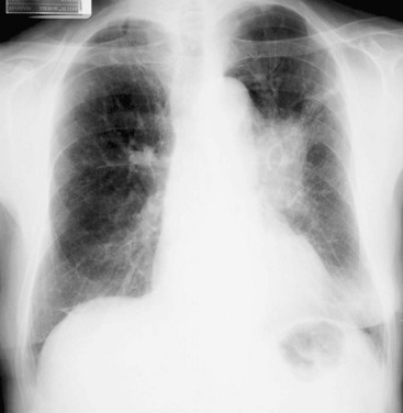
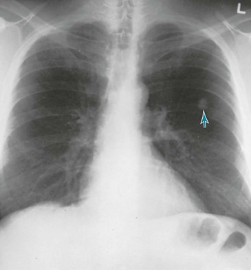
Lung Cancer: Clinical Aspects

21


Lung Cancer


Clinical Aspects


The goal of this chapter is to extend the discussion of lung cancer into the clinical realm and relate how the pathologic processes considered in Chapter 20 are encountered in a clinical setting. An outline of the major clinical features of lung cancer is followed by a discussion of the diagnostic approach and general principles of management. The chapter concludes with a brief discussion of bronchial carcinoid tumors, malignant mesothelioma, and the clinical problem of the solitary pulmonary nodule.


Clinical Features


Because lung cancer presumably starts with a single malignant cell, a long period of repetitive divisions and doubling of cell number must occur before the tumor becomes clinically apparent. During this preclinical period, an estimated 30 divisions take place before the tumor reaches a diameter of 1 cm. This process most likely requires a number of years, during which time the patient and physician are unaware of the tumor.


In general, the possibility of lung cancer is raised because of findings on imaging studies (chest radiography or computed tomography [CT]) or because of an assortment of symptoms that may ensue. This section focuses primarily on symptoms; imaging studies and diagnostic sampling are discussed under Diagnostic Approach. The symptoms at the time of presentation may relate to the primary lung lesion, to metastatic disease (either in intrathoracic lymph nodes or at distant sites), or to what are commonly called “paraneoplastic syndromes.”


Symptoms Relating to Primary Lung Lesion


Perhaps the most common symptoms associated with lung cancer are cough and hemoptysis. Because bronchogenic carcinoma generally develops in smokers, these patients often dismiss their symptoms (particularly cough) as routine complications of smoking and chronic bronchitis. With tumors originating in large airways (e.g., squamous cell carcinoma, small cell carcinoma), patients may also have problems related to bronchial obstruction, such as pneumonia behind the obstruction or shortness of breath secondary to occlusion of a major bronchus. In contrast, with tumors that arise in the periphery of the lung, including many adenocarcinomas and large cell carcinomas, patients tend not to have symptoms related to bronchial involvement, and their lesions are often found on a chest radiograph obtained for unrelated purposes. With all types of advanced lung cancers, constitutional symptoms such as malaise, anorexia, and weight loss occur frequently but are nonspecific.


When tumors involve the pleural surface, either by direct extension or metastatic spread, patients may have chest pain, often pleuritic in nature, or dyspnea resulting from substantial accumulation of pleural fluid. Other adjacent structures, particularly the heart and esophagus, can be involved by direct invasion or extrinsic compression by the tumor. Resulting complications include pericardial effusion, cardiac dysrhythmias, and dysphagia.


Tumors originating in the most apical portion of the lung, which are called superior sulcus or Pancoast tumors, often produce a characteristic constellation of symptoms and physical findings caused by direct extension to adjacent structures. Involvement of the nerves comprising the brachial plexus can result in pain and weakness of the shoulder and arm. Involvement of the cervical sympathetic chain produces the typical features of Horner syndrome—ptosis (drooping upper eyelid), miosis (constricted pupil), and anhidrosis (loss of sweat) over the forehead and face—all occurring on the same side as the lung mass. Invasion of neighboring bony structures (e.g., ribs, vertebrae) is a common complication.


Symptoms Relating to Nodal and Distant Metastasis


When the mediastinum has metastatic lymph nodes from a primary lung cancer, symptoms often arise from invasion or compression of important structures within the mediastinum, such as the phrenic nerve, recurrent laryngeal nerve, and superior vena cava. As a consequence, the following conditions, respectively, may develop: diaphragmatic paralysis (often with accompanying dyspnea), vocal cord paralysis (with hoarseness), and superior vena cava obstruction (with edema of the face and upper extremities resulting from obstruction to venous return).


Distant metastases, most commonly to the brain, bone or bone marrow, liver, and adrenal gland(s), frequently are asymptomatic. In other cases, symptoms depend on the particular organ system involved. Small cell carcinoma is the cell type most likely to generate distant metastases (see Chapter 20). Squamous cell carcinoma is least likely, and both adenocarcinoma and large cell carcinoma occupy an intermediate position.


Paraneoplastic Syndromes


Many lung tumors are capable of producing clinical syndromes not readily attributable to the space-occupying nature of the tumor or to direct invasion of other structures or organs. These syndromes are sometimes called the “paraneoplastic” manifestations of malignancy and frequently are due to production of a hormone or a hormonelike substance by the tumor. When a detectable hormone is produced by the lung tumor (or, for that matter, by any type of tumor), the patient is said to have “ectopic” hormone production. Sometimes clinical symptoms result from high circulating levels of the hormone; in other cases, only sensitive techniques of measurement are capable of demonstrating production of the hormone.


Why some tumors are capable of hormone production is not clear. Genetic information coding for the particular hormone is present but not expressed in the normal nonmalignant cell. In the course of becoming malignant, the cell presumably undergoes a process of gene dysregulation during which it regains the ability to express this normally silent genetic material coding for hormone production.


The cell type most frequently associated with ectopic production of humoral substances is small cell carcinoma, apparently because of its similarity or relationship to a type of neuroendocrine cell in the airway (the Kulchitsky cell) with secretory granules and the potential for peptide synthesis. Adrenocorticotropic hormone (ACTH) and antidiuretic hormone (ADH) are the best-described hormones produced by small cell carcinoma, potentially giving rise to the ectopic ACTH syndrome or to the syndrome of inappropriate ADH (SIADH), respectively. Squamous cell carcinoma is capable of causing hypercalcemia, which results from production of parathyroid hormone–related peptide, a peptide with parathyroid hormone–like activity. Production of other hormones, such as calcitonin and human chorionic gonadotropin, are also well described with bronchogenic carcinoma.


Other paraneoplastic syndromes cannot be attributed to a known hormone, and our understanding of their mechanisms varies. Examples range from a wide variety of neurologic syndromes (some of which appear to be due to autoimmune antibody production) to the soft tissue and bony manifestations of clubbing and hypertrophic osteoarthropathy (see Chapter 3). The nonspecific systemic effects of malignancy, such as anorexia, weight loss, and fatigue, are potential consequences of lung cancer, and it has been hypothesized that production of various mediators, such as tumor necrosis factor, may mediate these systemic effects.


Diagnostic Approach


A wide variety of diagnostic methods are used in evaluating cases of known or suspected lung cancer. Many studies assessing the lung on a macroscopic level aim to demonstrate the presence, location, and probability of spread of a bronchogenic carcinoma. Evaluation on a microscopic level is essential for defining the pathologic type of lung cancer, an important factor in determining what modalities of therapy are most appropriate. Functional assessment of the patient with lung cancer plays a role primarily in quantifying the severity of underlying lung disease, particularly chronic obstructive lung disease resulting from prior heavy smoking. Knowledge of a patient’s functional limitation from lung disease is essential before the clinician can decide whether operative removal of a lung cancer is even feasible without precipitating disabling respiratory insufficiency.


Macroscopic Evaluation


The initial test for detection and macroscopic evaluation of bronchogenic carcinoma is generally the chest radiograph. The presence of a nodule or mass within the lung on chest radiograph always raises the question of lung cancer, especially when the patient has a history of heavy smoking. The location of the lesion may give an indirect clue about its histology: peripheral lesions are more likely to be adenocarcinoma or large cell carcinoma, whereas central lesions are statistically more likely to be squamous cell carcinoma or small cell carcinoma (Figs. 21-1 and 21-2). The chest radiograph is also useful for determining the presence of additional suspicious lesions, such as a second primary tumor or metastatic spread from the original carcinoma. Involvement of hilar or mediastinal nodes or the pleura (with resulting pleural effusion) may be detected on the chest radiograph, and such a finding will substantially affect the overall approach to therapy.


CT has become a standard part of the diagnostic evaluation of patients with lung cancer. Besides helping to define the location, extent, and spread of tumor within the chest, this technique has been particularly useful for detecting enlarged, potentially malignant lymph nodes within the mediastinum, which are often not seen with conventional radiography. However, even though CT effectively identifies enlarged mediastinal nodes, it cannot definitively determine whether such nodes simply are hyperplastic or are enlarged because of tumor involvement. Consequently, histologic sampling of enlarged mediastinal nodes still is necessary to confirm tumor involvement of the nodes.


A newer technique that has gained increasing popularity in the evaluation of patients with known or suspected lung cancer is positron emission tomography (PET; see Chapter 3). Because of their high metabolic activity, malignant lesions typically exhibit high uptake of the radiolabeled glucose analog 18F-fluorodeoxyglucose (FDG). Focal uptake in the region of a parenchymal nodule or mass suggests the lesion is malignant, and uptake in the mediastinum or at distant sites often reflects spread of the tumor to those sites. However, other metabolically active lesions (e.g., focal areas of infection) may also show FDG uptake, so a positive PET scan in the appropriate clinical scenario is very suggestive but is not diagnostic of malignancy.


The best way to directly examine the airways of a patient with presumed or known bronchogenic carcinoma is by bronchoscopy with either a rigid or, much more frequently, a flexible bronchoscope (see Chapter 3). The location and intrabronchial extent of many tumors can be directly observed, and samples can be obtained from the lesion, either for cytologic or histopathologic examination. In addition, the bronchoscopist can assess whether an intrabronchial carcinoma is impinging significantly on the bronchial lumen and causing either partial or complete airway occlusion. Diagnostic specimens can be obtained in many cases even when the lesion is beyond direct visualization with the bronchoscope. In recent years, significant technologic advances in bronchoscopy, such as endobronchial ultrasound and electromagnetic navigation techniques, have increased the diagnostic yield of the procedure for lesions that are difficult to sample by direct visual or fluoroscopic guidance alone.


Staging of Lung Cancer


After a tumor has been documented, evaluation of the extent and spread (staging) of the malignancy is performed. In the case of non–small cell carcinoma of the lung (i.e., all cell types other than small cell carcinoma), staging is based on (1) the primary intrathoracic tumor—its size, location, and local complications, such as direct extension to adjacent structures or obstruction of the airway lumen; (2) the presence or absence of tumor within hilar and mediastinal lymph nodes; and (3) the distant spread of tumor to other tissues or organ systems. In the case of small cell carcinoma, the disease is classified as either limited (localized within one hemithorax) or extensive (beyond the limits of one hemithorax).


The first component of staging, taking into account characteristics of the primary tumor itself, is generally accomplished with a combination of chest radiography and bronchoscopy, sometimes with additional information obtained from CT or MRI.


The second component, based on involvement of mediastinal lymph nodes by tumor, is typically initially assessed by CT, sometimes complemented by PET. Definitive evaluation has generally been based on direct examination (and biopsy) of the nodes by either mediastinoscopy or mediastinotomy. In suprasternal mediastinoscopy, the mediastinum is visualized with a scope placed through an incision made just above the sternal notch. Biopsy specimens can be obtained by this technique if there is any suspicion that abnormal nodes are present. In parasternal mediastinotomy, the mediastinum is examined through a small incision made adjacent to the sternum, and samples of suspicious nodes can be taken. In selected cases, transbronchial needle aspiration

Stay updated, free articles. Join our Telegram channel

Jun 12, 2016 | Posted by in RESPIRATORY | Comments Off on Lung Cancer: Clinical Aspects

Full access? Get Clinical Tree

Get Clinical Tree app for offline access