Lipid Pneumonia
Timothy C. Allen MD, JD
Roberto Barrios MD
Lipid pneumonia, also termed endogenous lipoid pneumonia, postobstructive pneumonia, or “golden pneumonia,” occurs due to the escape of normally occurring lipids from lung tissue damage from a supparative process or from destroyed alveolar cell walls distal to an obstructing lesion such as lung cancer. It is termed “golden pneumonia” because affected lungs grossly have a golden or yellow-tan color because of the accumulation of lipid. Although large obstructions due to tumors are frequent, obstruction leading to lipid pneumonia may be so small as to involve only a small terminal or respiratory bronchiole. It is hypothesized that transbronchial dissemination of breakdown products from tumor cells, including mucin, may contribute to its spread in lung parenchyma.
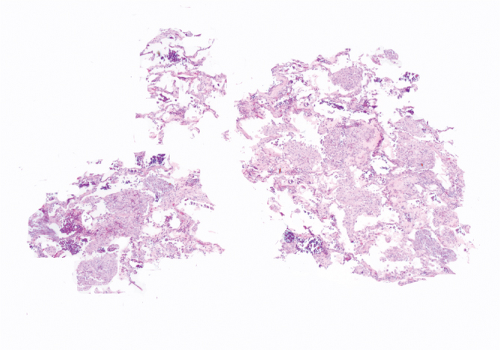
Histologically, there is accumulation of fine fat droplets in alveolar macrophages, with occasional associated giant cells and occasional cholesterol crystals. Scattered lymphocytes and plasma cells may be present and reactive type II pneumocyte hyperplasia. There is variable but generally mild interstitial widening with scattered chronic inflammatory cells and occasional interstitial foamy macrophages.
Differential diagnosis includes lipid storage diseases such as Gaucher disease and Niemann-Pick disease, lung reaction to amiodarone therapy, and lipoid pneumonia, also termed exogenous lipid pneumonia.
 Figure 23.1: Low power of transbronchial biopsy shows nodular aggregates of foamy cells consistent with lipid pneumonia.
Stay updated, free articles. Join our Telegram channel
Full access? Get Clinical Tree
 Get Clinical Tree app for offline access

|