CHAPTER 12 Left Lower Lobectomy—Video 12
 Video-Assisted Left Lower Lobectomy
Step 2: Inferior Pulmonary Ligament and Level 9 Lymph Nodes
♦ Aim the thoracoscope anteriorly, and point the 30-degree lens toward the inferior pulmonary ligament.
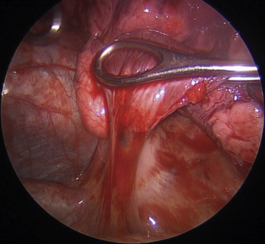
♦ Retract the lower lobe superiorly with a ring forceps through the utility incision. Apply enough tension to keep the inferior pulmonary ligament on stretch (Figure 12-1).
♦ Bring the cautery with an extension and the Yankauer suction into the chest through incision 1. If the diaphragm obstructs the view, push the diaphragm inferiorly with the Yankauer suction positioned so that its “bend” pushes the diaphragm downward while the tip is pointed toward the dissection to suck the smoke from the electrocautery.
♦ Alternatively, if the diaphragm is high and obstructing the view, retract it downward with a stitch. A needle holder through incision 1 places a suture in the ligamentous portion of the diaphragm, near the esophageal hiatus. Bring the suture with its needle out through incision 2. Pull it as tightly as possible and suture it to the chest wall inferior to incision 2.
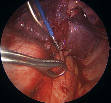
♦ Cauterize the inferior pulmonary ligament parallel to the esophagus up to the level of the inferior pulmonary vein (IPV) (Figure 12-2).
Step 3: Posterior Pleura
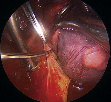
♦ With the lung retracted anteromedially, open the posterior pleura along the posterior surface of the IPV. Use a combination of sharp dissection with the Metzenbaum scissors and the long-tipped electrocautery, which are brought through anterior incision (Figure 12-3).
♦ The line of dissection posteriorly should be at the junction of the pleura and the lung parenchyma.
♦ In the course of this dissection, cut branches of the vagus nerve to the lung, and clip or cauterize bronchial arteries. Perform blunt dissection on the surface of the pericardium.
♦ Remove the lymph node on the posterosuperior aspect of the IPV. This is marked as a posterior hilar lymph node.


