Left Bundle Branch Block

3.4 Left Bundle Branch Block


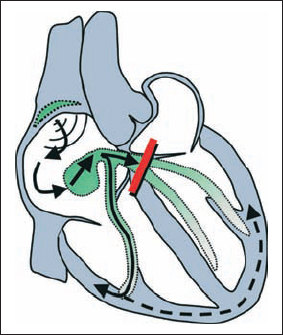
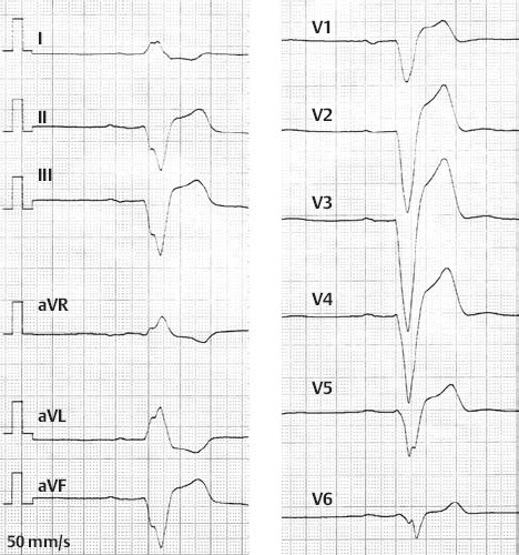
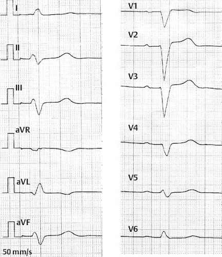
Isolated Complete Left Bundle Branch Block

image


Mechanism:


  • Block in the left bundle branch resulting in secondary (delayed) excitation of the left ventricle via the right ventricular myocardium

ECG characteristics:


  • QRS ≥ 0.12s; discordance of the ventricular repolarization (ST segment and T wave)
  • Ranging from left axis to marked left axis deviation
  • QRS configuration : V1/2 – “QS,” V5/6- “R’”
  • Turning point in V6: > 0.05 s


Etiology:


  • CHD, acute myocardial infarction (poor prognosis), heart defects, hypertension, medications that may cause conduction delay, carditis, DCM

Treatment:


  • Cessation of medications that may cause conduction delay
  • In symptomatic patients (syncope) bradycardia: insertion of pacemaker (or in cases of an unknown cause of syncope: electrophysiological study to exclude ventricular arrhythmias)
  • Conservative in the event of a lack of clinics

Isolated Complete Left Bundle Branch Block

image

image


Differential diagnosis:


In preexisting bundle branch block:


  • Atrial flutter with 3:1 conduction
  • AV conduction delay with SVES
  • 2nd or 3rd degree AV block
  • Prolonged P wave duration with interatrial block

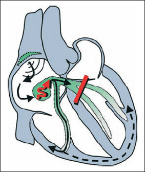
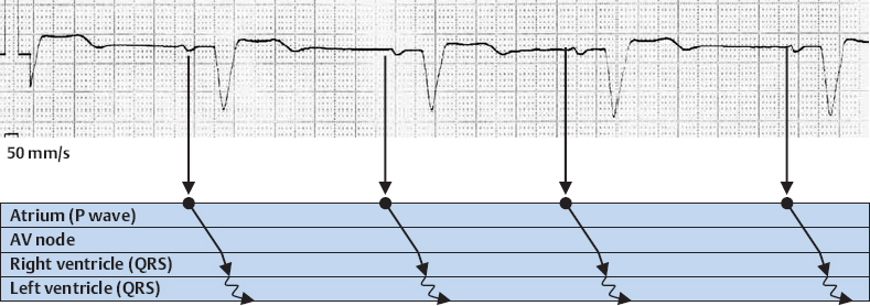
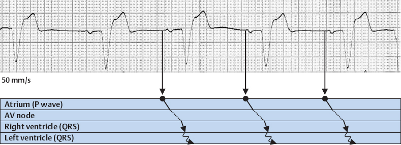
Complete Left Bundle Branch Block With 1st Degree AV Block

image


Mechanism:


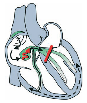
  • Delayed conduction at the AV node with additional block in the left bundle branch resulting in secondary (delayed) excitation of the left ventricle via the right ventricular myocardium

ECG characteristics:


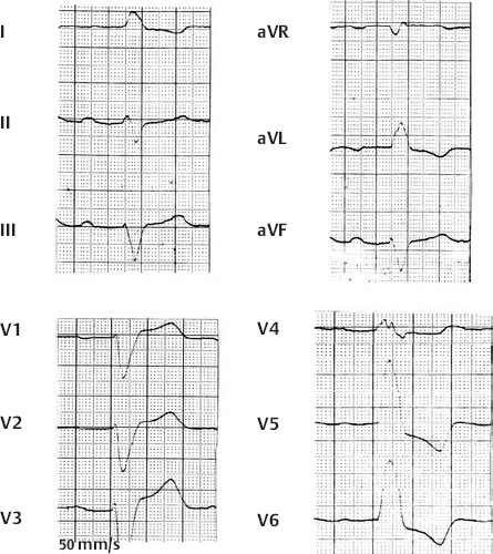
  • Complete left bundle branch block with delayed PQ interval (> 0.20 s)


Etiology:


  • CHD, acute myocardial infarction (poor prognosis), cardiac defects, hypertension, medications that may cause conduction delay, carditis, DCM

Treatment:


  • Cessation of medications that may cause conduction delay
  • In symptomatic patients (syncope) bradycardia: insertion of pacemaker (or in cases of an unknown cause of syncope: electrophysiological study to exclude ventricular arrhythmias)
  • Conservative in the event of a lack of clinics

Complete Left Bundle Branch Block With 1st Degree AV Block

image

image


Differential diagnosis:


In preexisting bundle branch block


  • Atrial flutter with 3 :1 conduction
  • AV conduction delay with SVES
  • 2nd or 3rd degree AV block
  • Prolonged P wave duration with interatrial block

Complete Left Bundle Branch Block With 1st Degree AV Block

image

image


Differential diagnosis:


In preexisting bundle branch block


  • Atrial flutter with 3:1 conduction
  • AV conduction delay with SVES
  • 2nd or 3rd degree AV block
  • Prolonged P wave duration with interatrial block

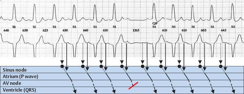
Complete Left Bundle Branch Block With 2nd Degree AV Block, Mobitz Type

image

Stay updated, free articles. Join our Telegram channel

Tags:
Aug 29, 2016 | Posted by in CARDIOLOGY | Comments Off on Left Bundle Branch Block

Full access? Get Clinical Tree

Get Clinical Tree app for offline access