Fig. 29.1
Retrogastric approach
At this point, approaching the MAL can be performed by three different techniques. The first technique is to follow the right crus of the diaphragm posteriorly to the left crus, which is the most commonly performed approach. The second technique is to start dissection from the left crus posteriorly to the right crus, and the third approach is to follow the branches of the celiac trunk back to the celiac artery until MAL is encountered.
The Right Crus Approach
This technique is the most commonly performed and has been described in several studies [7, 9, 12, 17, 18]. This technique was also the described technique in the pediatric MALS studies by Said et al. and Joyce et al. at the Mayo Clinic, Rochester, MN [17, 18].
Operative Technique
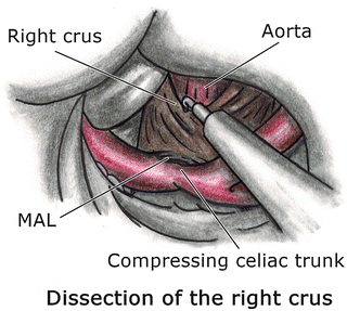
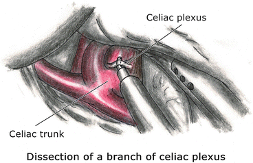
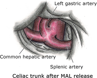
After the stomach is placed on left lateral traction, the right crus of the diaphragm is identified along with the caudate lobe of the liver and the inferior vena cava. The peritoneum over the right crus is opened using the L-hook electrocautery (Fig. 29.2). The esophagus and both the anterior and posterior vagal trunks are carefully identified. The retroesophageal space is then developed. This technique exposes the aorta first and then releases the median arcuate ligament cranially to caudally, directly anterior to the aorta coming onto the celiac artery. The decussation of the right and left crura is dissected to expose the aorta. Dissection proceeds caudally and must remain on the aorta anteriorly without veering off in either direction as there are no named branches until the celiac artery is encountered. The antrum of the stomach and the pancreas will likely need to be retracted caudally. Tight bands and ganglion fibers are lysed using the L-hook electrocautery on a low setting (Fig. 29.3). Because the fibers encountered here are tight and compress the celiac axis from anterior to posterior, these findings make the celiac axis nearly invisible. After one fiber at a time is lysed, the celiac artery will come into view as a bulge on the aorta (Fig. 29.4). Care must be taken not to back-heel (touch with the curved part of the cautery hook) any artery as one is lifting up on the individual fibers. Dissection is continued until the celiac, common hepatic, and left gastric arteries are free with 0.5–1.5 cm in either direction. One can consider re-approximating the crura cephalad to the MAL, though there is no compelling data to suggest that this be performed routinely.


Fig. 29.2
Dissection of the right crus

Fig. 29.3
Dissection of a branch of celiac plexus

Fig. 29.4
Celiac trunk after MAL release
The Left Crus Approach
This approach was described in a retrospective analysis of five consecutive patients who underwent laparoscopic division of the MAL by Nguyen et al. [19].
Operative Technique
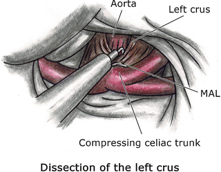
After the stomach is retracted superiorly and to the right, a liver retractor is placed under the posterior gastric wall. This technique is started by firstly approached the left crus (Fig. 29.5) and dissecting to the right crus, then to proceed dissecting the preaortic fibrous tissues as described above.


Fig. 29.5
Dissection of the left crus
The Vessel Approach
Some surgeons have found this approach to be more beneficial. Roseborough described this technique in his study. In several cases, final exposure of the origin of the celiac artery required retrograde dissection from the more distal celiac artery after first identifying the celiac axis or the hepatic artery distally [7]. A retrospective review by Kohn et al.. of the University of North Carolina, Chapel Hill, described this vascular dissection technique. A total of six patients were identified, two underwent a laparoscopic approach and experienced symptomatic improvement with a mean follow-up at 48.6 months [20]. Vaziri et al. also described this technique, by following the left gastric artery. They reported a case series of three patients with successful outcomes in a short-term follow-up period [13]. This technique was also described in the case reports by Wani et al. [21]
Operative Technique
This approach differs from the preceding description in the following steps: the avascular region of the gastrohepatic omentum (pars flaccida) is opened, and the left gastric artery identified. Using the L-hook electrocautery, dissection follows the left gastric artery and traces its origin up to the celiac trunk until the MAL is encountered, then proceeds through dissecting the preaortic fibrous tissues, celiac plexus, and the MAL as described above.
Antegastric Approach (Gastrohepatic Ligament/Pars Flaccida Approach)
Because it is similar to the technique in gastroesophageal operations such as laparoscopic Nissen fundoplication, this approach for the MAL release has been described and performed by many institutions [14]. A retrospective study by Tulloch et al.. described this approach combined with the right crus technique and demonstrated a technical success rate in the laparoscopic technique group of 80 %. Twenty percent required conversion for bleeding. Operative conversion was required for acute bleeding from the left gastric artery in one patient and the celiac trunk in the other. Both open conversions occurred early in our experience and likely occurred due to subadventitial dissection and excessive thinning of the artery with the ultrasonic dissection. Direct suture repair of the left gastric artery was performed during the first conversion, and patch angioplasty was required to repair the celiac trunk arteriotomy during the second [9].
Operative Technique
The avascular region of the gastrohepatic omentum, also known as the pars flaccida, is identified. A grasper is used on the stomach, retracting it inferiorly and laterally such that tension is placed on the gastrohepatic ligament. An ultrasonic dissector or another cutting/sealing device is used to open the gastrohepatic ligament (Fig. 29.6). The esophagus is dissected circumferentially, exposing the right crus of the diaphragm. The right crus is then isolated inferiorly to the cardia. A Penrose drain or a vascular loop is used to retract the esophagus and/or stomach to gain better exposure of the aortoceliac region. Once the crura of the diaphragm are separated, the abdominal aorta is identified. The preaortic fibrous tissues are divided using the “L”-hook electrocautery as described in the right crus technique. The other two techniques (following left crus and the branches of celiac artery) can also be used. The MAL release is considered complete once the celiac axis is clearly visualized without any residual impinging tissues. This generally results in a visible change in the trunk’s position and orientation, albeit sometimes subtle. The trunk has been described as “rising” off the aorta by a small amount.


Fig. 29.6
Antegastric approach
The Robot-Assisted Technique for MAL Release
Jaik et al. first reported the successful treatment of MALS utilizing a robotic surgical system [6]. Subsequent reports by Antoniou et al. and Meyer et al. also describe the technique of robotic approach for division of the MAL [22, 23]. This technique was also described in a retrospective review performed by Do et al.. from the Ochsner Clinic Foundation, New Orleans, LA. Comparing the outcomes between laparoscopic and robot-assisted treatment of MALS, a total of 16 patients, 8 out of 12 patients (67 %) in the laparoscopic group and 2 out of 4 patients (50 %) in the robot-assisted approach group, reported complete resolution of their abdominal pain in the 20-month follow-up. No intraoperative or perioperative complications nor conversions were reported, though the study size was obviously quite small [24].
In institutions that employ the surgical robotic system, the benefits of robotic surgery may be due to the stereoscopic view of the surgical field, which enhances depth perception, and the improved surgical precision of movement due to the wrist-like articulations of the instruments, enhancing performance in the hard-to-reach areas. This is most obvious if the patient has a somewhat high-riding pancreas, such that the superior border of the pancreas is crowding the area of dissection. The maneuvering around the celiac artery has shown to be improved in terms of the dexterity with the use of the enhanced three-dimensional display inherent to the robotic system when compared to monoscopic laparoscopic systems [10]. However, disadvantages include higher cost and increased operative time and time for equipment setup. Operative time was 168 min in the reported case, in contrast with a mean of 90 min for the laparoscopic technique [12].
Operative Technique
Once a liver retractor is placed exposing the anterior gastric wall, approaching the lesser sac can be performed either with the retrogastric or the antegastric approach.
The robotic system is docked, and meticulous dissection is undertaken in the region of the confluence of the left gastric artery and the hepatic artery. Dissection in this area is carried down until the crura are exposed at the base where the left crus crosses the right crus. In the antegastric approach, the stomach is retracted to the left to gain better exposure. Subsequently, the left gastric artery can be looped with a Penrose drain and also retracted to the left. The right crus dissection is carried out caudally. Upon further dissection, the aorta and celiac artery come into view and the right diaphragmatic crus is seen coursing over the aorta and around the celiac axis. The right crus is then carefully divided. All the fibrous attachments around the aorta and the origin of the celiac artery are meticulously dissected with the L-hook electrocautery along with the celiac nerve plexus and lymphatic tissue. Division of these fibers on the celiac artery is carried out for some distance onto the celiac artery circumferentially until the artery appears to be free of any external compression. Once these maneuvers are completed, the celiac axis will be clearly visualized without any residual kinking and uniform throughout its course.
Stay updated, free articles. Join our Telegram channel

Full access? Get Clinical Tree


