Kawasaki’s Disease

56 Kawasaki’s Disease


Kawasaki’s disease is a multisystem vasculitis, first recognized in 1961 and subsequently described in 1967 by Dr. Tomisaku Kawasaki. Kawasaki’s disease was initially thought to be a benign, self-limited febrile illness. The association of the disease with the development of coronary aneurysms and subsequent mortality did not become evident until the 1970s. Since then, Kawasaki’s disease has been recognized in all populations and is now reported to be the most common acquired heart disease in U.S. children, replacing rheumatic heart disease.


The incidence of Kawasaki’s disease varies among ethnic groups. It is highest in children of Asian ancestry (90 per 100,000 in Japan) and lowest in certain Europeans (3 per 100,000 in Britain). The U.S. incidence is 17 per 100,000 in African-Americans, 11 per 100,000 in Hispanics, and 9 per 100,000 in Caucasians. Kawasaki’s disease usually occurs in children 1 to 2 years of age, with 80% of cases diagnosed in children younger than 4 years of age. However, the diagnosis has been made in children of all ages. Males are more commonly affected than females (a ratio of 1.5 : 1).


Etiology and Pathogenesis


The etiology of Kawasaki’s disease is unknown. A genetic predisposition to the disease is suggested by the higher incidence in children of Asian ancestry and in siblings. The occurrence of the disease in siblings also suggests exposure to a common causative agent. Various bacteria, viruses, heavy metals, and detergents have come under suspicion as etiologic agents. A superantigen (toxin)-mediated hypothesis has been proposed, but large multicenter studies have failed to conclusively identify any single etiologic agent or group.


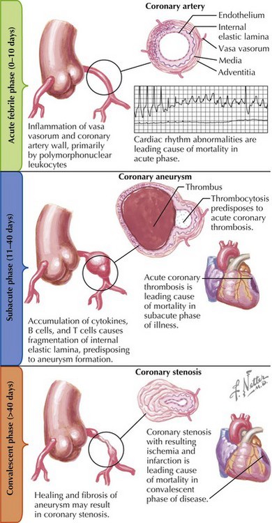
Kawasaki’s disease is a nonspecific vasculitis that affects small- and medium-sized arteries throughout the body. Its most serious consequences evolve in phases within the coronary arteries (Fig. 56-1).


The acute febrile phase of Kawasaki’s disease may last less than a day or up to 10 days. Inflammation in the coronary artery walls consisting mostly of polymorphonuclear cells may occur. An increased ratio of T4 (helper) to T8 (suppressor) cells has been found in vessel walls during the acute phase. Pancarditis may also develop during the acute phase. However, the mortality rate during this phase is low, and death is thought to occur only in severely affected individuals as a result of myocardial dysfunction or arrhythmias.


The subacute phase lasts 10 to 40 days. An accumulation of cytokines, B cells, and T cells in the vessel walls may cause fragmentation of the internal elastic lamina of the coronary arteries and, subsequently, formation of an aneurysm. Thrombocytosis occurs simultaneously and predisposes patients to acute coronary thrombosis, the leading cause of mortality during this phase of the illness.


The convalescent phase (beyond 40 days) consists of healing and fibrosis of the coronary aneurysms formed during the subacute phase. A stenosis may occur at such sites, with resulting ischemia, infarction, and death. Of all deaths, 70% occur in infants younger than 1 year of age, and almost all deaths occur during the convalescent phase.


The risk factors for the development of coronary artery disease include fever lasting more than 14 days, recurrence of fever or rash, treatment with intravenous immunoglobulin (IVIG) more than 10 days into the acute illness, male sex, and age younger than 1 year.


Clinical Presentation


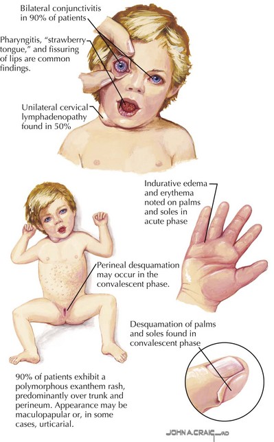
Fever is the cardinal presenting sign of Kawasaki’s disease and persists for more than 7 days in more than 95% of patients. It responds only temporarily to antipyretic therapy and may last up to 2 weeks in patients who are not treated with IVIG. Within 5 days of the onset of fever, 90% of patients develop a polymorphous rash predominantly over the trunk (Fig. 56-2). This may be a diffuse maculopapular rash, or it may be a more urticarial rash. Rarely, perineal desquamation is seen in the subacute phase. Mucosal changes, erythema, and fissuring of the lips occur in 90% of patients. Pharyngitis and prominent papillae (“strawberry tongue”) are also typical signs. Within the first week of the illness, bilateral conjunctivitis, consisting of a discrete vascular injection with absence of corneal clouding or purulent exudates, develops in 90% of patients. The bilateral conjunctivitis persists for 1 to 2 weeks. Anterior uveitis is a common finding on slit-lamp examination. Indurative edema and erythema of the hands and feet occur in 75% to 90% of patients. Desquamation of the fingertips and/or the palms and the soles may occur 2 to 3 weeks after the acute illness. Cervical lymphadenopathy, the least consistent finding, occurs in approximately 50% of patients. It is usually unilateral, and the diameter of the nodes may exceed 15 mm.


Stay updated, free articles. Join our Telegram channel

Jun 12, 2016 | Posted by in CARDIOLOGY | Comments Off on Kawasaki’s Disease

Full access? Get Clinical Tree

Get Clinical Tree app for offline access