Fig. 14.1
(a, b, c) Serial chest radiographs showing a relatively normal cardiothoracic ratio and pulmonary venous congestion
It was mid-October and the beginning of annual winter bronchiolitis epidemic in the UK. She was reviewed by a Consultant Paediatrician and a diagnosis of bronchiolitis was made. She was managed with headbox oxygen, antibiotics and continuation of breast feeds. Over the next 5 h she became increasingly tachypnoeic (respiratory rate 80 and 100 breaths/min) and tachycardic (heart rate 160–170/min). During a breast-feed she suddenly became pale and dropped her oxygen saturations to 40 %. Trial of continuous positive airway pressure (CPAP) failed; she was therefore intubated and ventilated and transferred to the Paediatric Intensive Care Unit (PICU).
On admission to PICU, she was noted to have normal heart sounds, no murmurs and good volume femoral pulses with a heart rate of 140–150/min and a blood pressure of 70/55 mmHg. Auscultation of chest revealed bilateral equal air entry with no additional wheeze or crackles. Liver was palpable 4 cm below the right costal margin. Endotracheal suction yielded thick white secretions. She was afebrile and her admission CRP was <4, with slightly elevated WCC (17.6 × 10⁹/l) and neutrophil count (9.5 × 10⁹/l). Radiology again reported the chest radiograph as showing ‘bilateral perihilar ground glass appearance with streaky opacification consistent with bronchiolitis’ (Fig. 14.1b).
During her stay, she required moderate ventilatory pressures (PIP 24, PEEP 6, rate 35/min) and the FiO2 decreased from 60 % to 35 % maintaining her oxygen saturations greater than 95 %. Her arterial blood gases were satisfactory (pH 7.44, PCO2 4.12 kPa, PO2 9.2 kPa, BE R-3, Lactate 0.4). She was sedated and paralysed with morphine and rocuronium infusions. She passed an adequate volume of urine and nasogastric feeds were well tolerated. All these features reassured the team that the clinical diagnosis was most likely bronchiolitis. She did, however, become significantly bradycardic and hypoxic with any stimulation, such as suction or changing her position. It was believed that these episodes could be due to vagal stimulation, and the endotracheal tube was changed from oral to nasal in an attempt to resolve this issue.
Over the following 48 h, her ventilator settings remained more or less the same. However, she continued to have episodes of bradycardia, with a mottled appearance and profound desaturations during any intervention. Hyperoxia test (100 % FiO2 for 10 min) yielded a PaO2 of 31 kPa. She continued to have thick white secretions requiring regular saline washouts. In the meantime her respiratory secretions did not grow any bacteria or viruses. Repeat chest radiograph showed increased airspace shadowing, especially on the right side (Fig. 14.1c).
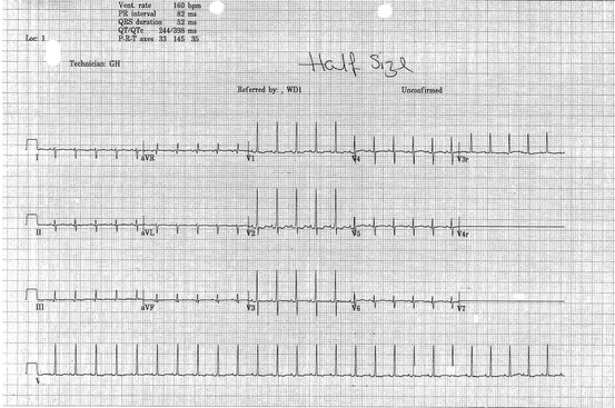
In view of negative microbiology with low inflammatory markers, persistent desaturations with handling, static clinical course and worsening chest x-ray picture, an echocardiogram was requested on day 3 of admission. This showed infracardiac total anomalous pulmonary venous connection (TAPVC) with no obvious obstruction and an atrial septal defect (ASD) with right to left flow. A 12-lead ECG showed normal sinus rhythm, right axis deviation (QRS + 120), tall R-waves in V1/V2 and inverted T wave, consistent with right ventricular hypertrophy (Fig. 14.2).


Fig. 14.2
Twelve-lead ECG, half standard, illustrating right ventricular hypertrophy
Following the diagnosis, she was referred to the local cardiology centre, and arrangements made for transfer. In the next 24 h, she deteriorated and behaved as though the pulmonary veins were obstructed, requiring increasing respiratory and cardiovascular support. She was transferred to the cardiac surgical centre on the fourth day following her admission to hospital. A CT scan was performed to define the pulmonary venous anatomy and confirmed infracardiac TAPVC.
She was operated 5 days after her initial presentation to hospital. Intraoperative findings confirmed an infracardiac TAPVC, with a vertical connecting vein formed by the confluence of right and left pulmonary veins. This connecting vein drained to inferior vena cava via the portal vein. An anastomosis was performed between the connecting vein and left atrium, and the ASD was closed. Her post-operative period was complicated with recurrent pleural effusions. She was eventually discharged home 1 month after her operation. Follow-up 6 weeks later revealed a thriving baby who was feeding and growing well. Repeat echocardiogram confirmed a good surgical result with a widely patent pulmonary venous anastomosis. She is now 2½ years old and is doing well.
Discussion
Total anomalous pulmonary venous connection (TAPVC) is a rare form of congenital heart disease in which all pulmonary veins connect to the systemic veins, right atrium or coronary sinus. In order to survive, there is an obligate interatrial communication. TAPVC can occur in conjunction with a wide variety of other cardiac malformations, in particular atrial isomerism. The disease was first described by Wilson in 1798.
< div class='tao-gold-member'>
Only gold members can continue reading. Log In or Register to continue
Stay updated, free articles. Join our Telegram channel

Full access? Get Clinical Tree


