Fig. 4.1
Brain axial CT showing left temporal focal bleeding
Vital Signs
Heart rate: 80 bpm
Blood pressure: 108/80 mmHg
Oxygen saturation: 98 %
Physical Examination
General: traumatic scalp wound 4 cm
Neurological: mental status was impaired. Pupil exam showed pupil asymmetry <1 mm and no dilatation or constriction; pupils were reactive to light and accommodation bilaterally. No alterations in deep tendon reflexes.
Cardiovascular: regular rate and rhythm, S1 and S2 normal, no murmurs, rubs, or gallops, and no hepatojugular reflux.
Lungs: no signs of respiratory effort on inspection. No wheezes, no crackles or stridor, and no bronchial or vesicular breath sounds on auscultation over the lung fields bilaterally.
Abdomen: no lesions or signs of trauma. No pain or rebound on light and deep palpation, no organomegaly, and normal bowel sounds in all four quadrants.
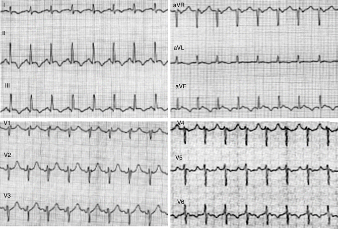
Routine ECG was performed after the hospitalization, showing sinus rhythm, normal atrioventricular conduction, incomplete right bundle branch (RBBB), and no alterations in repolarization.
Laboratory Exams on Admission and Other Tests
Exams (including high-sensitivity cardiac troponin) were unremarkable except for a mild leukocytosis of 14,500/mm3.
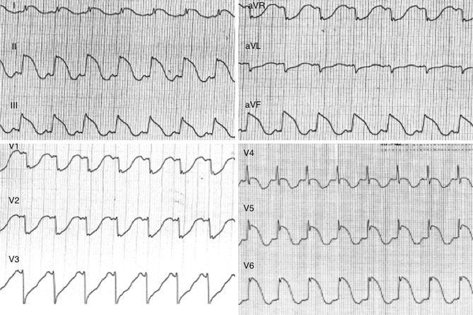
After 2 days in the hospital, the patient had a control CT of the brain that showed an increased size of the left temporal subarachnoid hemorrhage, with minimum brain compression and appearance of posterior subcortical-cortical bleeding. The patient was clinically stable, but ECG changed dramatically and showed sinus tachycardia; normal atrioventricular conduction; ST elevation in DII, DIII, aVF, V4, V5, and V6; and ST depression in aVR, aVL, V1, V2, and V3 (Fig. 4.2).


Fig. 4.2
Rest ECG showing incomplete right bundle block and normal repolarization
Fast echocardiography was performed, revealing no cardiac contractile abnormalities. Blood sample showed a rise in troponin level: 0.16 ng/ml.
What Are the Possible Causes for ST Elevation?
Acute ST-segment elevation myocardial infarction (STEMI)
Coronary spasm
Pericarditis
Left ventricular aneurysm
Brugada syndrome
Left bundle branch block
Left ventricular hypertrophy
Early repolarization
Drugs (e.g., cocaine, digoxin, quinidine, tricyclics, and many others)
Electrolyte abnormalities (e.g., hyperkalemia)
Neurogenic factors (e.g., stroke, hemorrhage, trauma, tumor, etc.)
Metabolic factors (e.g., hypothermia, hypoglycemia, hyperventilation)
What Are the Possible Causes of Increased Troponin Level?
Acute coronary syndrome
High blood pressure in lung arteries (pulmonary hypertension)
Blockage of a lung artery by a blood clot, fat, or tumor cells (pulmonary embolus)
Congestive heart failure
Myocarditis
Prolonged exercise
Trauma that injures the heart
Weakening of the heart muscle (cardiomyopathy)
Long-term kidney disease
In a setting of acute coronary syndrome and ST elevation, the patient has been promptly treated with metoprolol and acetylsalicylic acid intravenous and a percutaneous coronary angiography was urgently performed which showed just a non-critical stenosis (<30 %) of the right coronary artery. In the following 48 h, ST elevation gradually resolved, and waves were inverted in DII, DIII, aVF, V4, V5, and V6 (Fig. 4.3). A repeated head CT scan revealed a partial reduction of the encephalic bleeding. The patient was extubated and transferred to a spoke intensive care unit.
 < div class='tao-gold-member'>


Only gold members can continue reading. Log In or Register to continue
Stay updated, free articles. Join our Telegram channel

Full access? Get Clinical Tree


