Intestinal Bleeding and Atrioventricular Block
A 72-year-old woman was hospitalized due to intestinal bleeding. Her electrocardiogram showed a first-degree atrioventricular block. She had no dyspnea or chest pain and occasional palpitations. See Videos 66-1 and 66-2 and Figures 66-1 and 66-2.
An additional contrast study with agitated saline was performed. In Videos 66-3 and 66-4 and Figures 66-3 and 66-4, the contrast agent is applied once via a right cubital vein and once again via a left cubital vein.
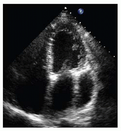 Figure 66-1. Transthoracic echocardiogram (TTE): Apical four-chamber view. |
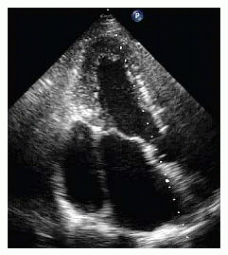 Figure 66-2. TTE: Apical three-chamber view. |
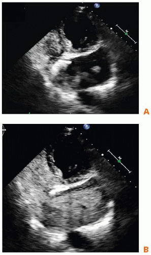 Figure 66-3A,B. TTE: Atypical apical four-chamber view. Agitated saline is injected into the right cubital vein. The agitated saline appears in the right atrium first, then the coronary sinus is opacificated. |
QUESTION 1. What is the patient’s diagnosis?
A. Coronary sinus aortic stenosis
B. Circumscribed pericardial effusion
C. Cor triatriatum sinister
D. Cor triatriatum dexter
E. Persistent left superior vena cava (PLSVC)
View Answer


ANSWER 1: E. The patient has a PLSVC, which is considered the most common abnormality of the thoracic venous system.
Stay updated, free articles. Join our Telegram channel
Full access? Get Clinical Tree
