Fig. 10.1
A 3-D reconstruction from CT angiogram of the abdomen and pelvis
10.1.2 Angiography
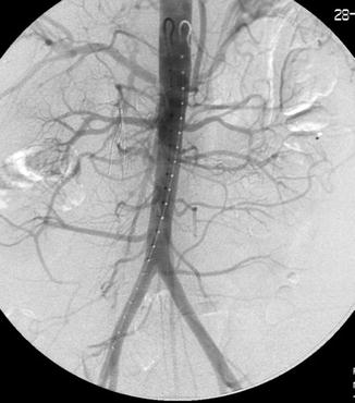
The majority of angiography is currently performed using digital subtraction. Digital subtraction angiography (DSA) provides visualization of blood vessels by subtracting out background structures before contrast injection. DSA can be performed with multi-station or stepping table (bolus chase) techniques. Multi-station DSA is performed in sections, each with its own injection (e.g., upper arm, lower arm, hand). This usually gives the best images particularly for peripheral angiography but increases radiation dose and contrast administration. Stepping table DSA is used only in peripheral angiography. The C-arm or table moves in a series of overlapping steps with a single bolus of contrast injection. This technique usually requires a more experienced technician and is of limited utility except in bilateral lower extremity pathology (Fig. 10.2).


Fig. 10.2
Digital subtraction angiography (DSA) of the abdomen and pelvis (AP view)
10.2 Equipment
10.2.1 Fluoroscopy
Most modern angiography units use pulsed fluoroscopy where radiation is produced in a pulsating fashion instead of a continuous beam. The pulses typically range from 2 to 30 pulses per second. The primary advantage of pulsed fluoroscopy is a significant reduction in radiation dose. The goal of fluoroscopy is to hold the radiation exposure to both patient and operator to a minimum while still providing the image detail necessary for the given situation. In addition to the use of pulsed fluoroscopy, this can be achieved by keeping the image intensifier or flat-panel detector close to the patient, centering over the region of interest, judicious use of collimation, moving the table before screening when changing positions, and optimizing single angiographic runs to sort out anatomy. Judicious application of fluoroscopy and features such as “last image hold” where the last image is displayed on the monitor also help reduce radiation exposure to the patient, technologists, nurses, and physicians.
10.2.2 Power Injectors
Power injectors deliver a controlled bolus of contrast. They have significant advantages over hand injections by delivering contrast boluses with higher flow rates, consistent volumes, and precise timing. Parameters settings include flow rate (ml/s), total volume (ml), pressure rate rise, linear rise, maximum pressure (psi), and injection delay. Flow rate is selected on the basis of vessel diameter and blood flow. Volume depends on the desired column length and size of area of interest. Pressure rate rise is the time to peak pressure and typically set at 0.4 s. Although most modern angiographic catheters have very high pressure tolerance, catheter-specific maximum pressure is set to avoid any chance of catheter rupture. The pressure generated by a power contrast injection is dependent on contrast viscosity, flow rate, catheter luminal diameter, and catheter material and length. Linear rise is the time required to reach the maximum flow rate. It is used to avoid catheter recoil and potential vessel injury. Rise time depends on catheter size and position. For example, a linear rise of 0.2 s is typical for multi-side hole catheters but a longer time (0.4–0.8 s) may be required for an end-hole catheter. Injection delay can be variable depending on the distance of the area of interest from the catheter delivering the contrast bolus.
10.2.3 Vascular Access Sheaths
When bleeding is suspected, vascular access must be gained by an artery. Typically this is the femoral artery, although brachial or even radial arterial access methods have been employed. Vascular sheaths provide an atraumatic conduit for vascular access and are particularly helpful when multiple catheter exchanges are required. By convention, a sheath size is rated for size of the catheter it will accept in its lumen but has a larger outside diameter. For example, a 5 Fr sheath will accept a 5 Fr catheter (1 “French” equals approximately 0.33 mm) but produces a 6.5 Fr hole in the artery. The sheath has a hemostatic valve where catheters are inserted and a sidearm for flushing to which a “flush bag” can be attached to prevent thrombus formation at the tip of the sheath.
10.2.4 Guidewires
Guidewires serve to negotiate a pathway from entry access to target area. Along with the choice of an appropriate catheter, the guidewire used is the key element in the success of any selective or superselective catheterization and ultimately the success or failure of any diagnostic or interventional procedure. Guidewires are divided into two major categories: non-steerable and steerable. Non-steerable guidewires provide a rail for catheters to be advanced into crude position but not navigate into select branch vessels. Typical examples of non-steerable guidewires include the following: J guidewire (most common), Straight, Bentson, Rosen, Amplatz, and Lunderquist. Steerable guidewires have shaped or shapeable angled tips allowing them to be selectively rotated and directed through tight stenoses or areas of tortuosity into branch vessels.
Selection of guidewire application is further based on diameter, length, material (e.g., stainless steel or nitinol), lubricity (hydrophilic or not), wire tip shape, and overall stiffness. Most guidewires are either microwires (0.010–0.018 in.) or standard (0.025–0.038 in.). The majority of cases start with use of a standard 0.035 in. guidewire which will fit through 4 or 5 Fr catheter. Small caliber wires (typically 0.010–0.018 in.) are used with microcatheters, usually coaxially through a 5 Fr base catheter. Length of the guidewire is critical when considering catheter exchanges to gain favorable position. A standard guidewire is 145–180 cm, but longer wires may be required for working from groin to upper limb or visceral circulation. Wire tips are floppy and atraumatic at one end and rigid at the other end. The length of the floppy tip can vary and application is based on optimizing the transition zone (floppy to stiff portion) within the target vessel. Wire stiffness is a key criterion in selection of non-steerable wires. Stiffness choice is made based on task required. For example, a 5 Fr diagnostic catheter can be advanced over a standard guidewire, but, depending on body habitus or vascular tortuosity, a large sheath or guiding catheter used to deliver a stent may require the use of a stiffer guidewire. Very flexible microwires with shapeable tips are often required to select small vessels, over which a microcatheter can be positioned for superselective embolization.
10.2.5 Catheters
Like guidewires, catheters are divided into two categories: nonselective (diagnostic) and selective. Catheters are named for their outer lumen size (e.g., 5 French). Nonselective catheters, also known as flush catheters, are used to inject medium to large-sized vessels and have multiple side holes to deliver large boluses of contrast. A pigtail catheter is the most commonly used nonselective catheter. Pigtail catheters are marketed in a variety of iterations and have both end and side holes which provide largest flow at the end with homogenous contrast bolus from the side holes. Maximum flow rate of catheters depend on luminal diameter, length, and number of side holes. The maximum injection rate and injection pressure is listed packaging and catheter hub.
Selective catheters have a wide variety of shapes and are used to selectively access branch vessels. Selective catheters may have end hole only or end and side holes. End-hole catheters should not be used indiscriminately with power injectors due to the high flow jet created which may lead to possible vessel injury or dissection. Catheters are further classified by their material (e.g., hydrophilic) and their composition, braided or non-braided. A braided catheter has a small amount of metallic fiber woven into the walls which makes the catheter more steerable and improves trackability.
Common selective catheters include Cobra, Berenstein, Sidewinder, and Headhunter. Each of these catheters is designed for specific target regions such as head and neck, aortic arch, visceral, and peripheral. Most cases start with a diagnostic catheter (e.g., 5 Fr pigtail), and then selective catheters are chosen based on the intended treatment vessel, but changes are often made during a case based on the given anatomy. A microcatheter is employed coaxially for more distal access into smaller vessels where advancing larger catheters might be difficult or impossible, might be occlusive, or cause vessel spasm or injury (Table 10.1).
Table 10.1
Measurement standard nomenclature for angiography catheters, wires, and sheaths
French sizes (circumference mm) | Inches | Centimeters |
|---|---|---|
Angiographic catheter (outer) | Wire diameter | Wire length |
Guide-catheter size (outer) | Catheter lumen | Catheter length |
Sheath size (inner) | Sheath length |
10.3 Interventional Technique
It must be noted that while techniques for access, vascular catheterization, and endovascular treatment are described in this chapter, these types of interventions should not be performed without first having received direct training and guidance by an experienced endovascular interventionalist.
10.3.1 Vascular Access
Access puncture sites are fixed and compressible over bone for hemostasis. The common femoral artery (CFA) is the most widely used site because of the size of the vessel and lowest risk of complications at the site. Brachial access is occasionally employed but even small amount of hemorrhage at this site carries potential adjacent nerve damage. The choice of ipsilateral or contralateral, or antegrade or retrograde femoral access for lower extremity arteriography or intervention depends on the index of suspicion of injury, intended treatment plan, and age and clinical status of the patient. For example, one might not want to access the right common femoral artery in an individual with a right acetabular fracture. The elderly often have markedly tortuous iliac vasculature that might make contralateral catheterization difficult, and marked obesity might make safe antegrade femoral access difficult or impossible.
The CFA courses over the medial half of the femoral head. Pulse is typically used as the landmark, but position of the femoral head should be checked with fluoroscopy as the inguinal crease is not a reliable landmark, particularly in obese patients. Ultrasound-guided puncture can facilitate arterial access in difficult cases or may even be utilized on a routine basis.
It is recommended that puncture of the femoral artery should be at the level of the midpoint of the femoral head. The skin entry site and course of the needle should account for this such that a more inferior skin entry will be required. Puncturing the CFA too high (above the inguinal ligament) increases the risk of bleeding due to inability to compress the artery after sheath removal. Too low of a puncture carries similar risk but may lead to occlusion of the superficial femoral artery. Access of the CFA is typically performed either with a micropuncture set or standard vascular access kit. The micropuncture set uses a smaller gauge needle (21 G) and accepts a 0.018-in. wire with floppy tip. Once the vessel is accessed with the 21 G needle, the 0.018-in. wire is passed, over which a two-part dilator is introduced. The 3 Fr inner component accepts and accommodates the small 0.018 in., and the outer 4 Fr component has a 0.035-in. diameter. Once the inner component is removed, a standard 0.035-in. guidewire can be passed and allow the placement of a vascular sheath. The standard access kit uses an 18/19 G needle that will accept a 0.035-in. guidewire. The micropuncture set has the advantage of securing an optimal puncture with less risk of trauma to the vessel.
10.3.2 Sheath Placement/Passage of Guidewire
Access of the artery with a micropuncture set as described above is recommended. With the 21 G needle in place and pulsatile blood return confirmed, the 0.018-in. wire should be slowly advanced under fluoroscopic guidance. The needle position over the femoral head can be confirmed with the wire in place. If too high or too low, withdraw the needle and wire, apply pressure for several minutes, and try again. When manipulating the wire, the tip of the wire should be in view at all times. If there is any resistance, withdraw a few centimeters, and then advance again looking for deviation from the anticipated path. If the 0.018-in. wire will not advance intraluminally, it is likely the needle tip has been advanced too far into the vessel wall. Withdraw the wire, check for pulsatile flow, and reposition the needle if necessary. If the wire is intraluminal and will not advance, ensure that the wire is taking the anticipated path and not in a branch vessel. If redirection is not successful, a 4 Fr dilator can be inserted to secure the access. Make certain that the dilator can be aspirated. Gently inject contrast to confirm intraluminal position then hand-inject an angiogram to identify the problem. Once access is established with the 4 Fr micropuncture set, the inner portion and 0.018-in. wire are removed and a 0.035-in. guidewire is advanced through the lumen. A J-wire with a very floppy tip (e.g., LLT or Benson) is the most commonly used starter guidewire. Once this wire is in place, typically a 5 Fr 10-cm sheath is placed over the wire. Use of steerable hydrophilic wires or shaped catheters may be necessary to navigate diseased or tortuous vessels. Always ensure a suitable length of the guidewire in the vessel before introducing a sheath/dilator or exchanging a catheter.
10.3.3 Introduction of Catheter
Catheter selection is dependent on the procedure. The catheter should be move easily over a guidewire which must be fixed and under tension (i.e., the system must be kept straightened). Always insert an ample amount of guidewire under fluoroscopic visualization into the vessels before passing the catheter. The wire leads the catheter into a large vessel. The wire is then advanced to the target followed by the catheter placement into the branch vessel for angiography or intervention. Catheter shape, in addition to steerable guidewires, will be the main determinant of the ability to successfully catheterize the intended target vessel. Most microcatheters have radiopaque tips to allow easy identification of the catheter tip. A small hand injection of contrast under fluoroscopy should always be performed to gauge catheter position and blood flow before DSA is performed.
Stay updated, free articles. Join our Telegram channel

Full access? Get Clinical Tree


