Study
N
Population
Study question
Comparison
Findings
Fischer
189
Resectable stage I-III NSCLC
Number of ‘futile thoracotomies’
CS - > S
52 % vs. 35 % (P = 0.05)
PET-CT - > S
Maziak
337
Resectable stage I-IIIA NSCLC
Proportion in whom correct upstaging
CS - > S
7 % vs. 14 % (P = 0.046)
PET-CT - > S
Ung
310
Unresectable stage III NSCLC
Proportion in whom correct upstaging
CS - > RT
3 % vs. 15 % (P = 0.0002)
PET-CT - > RT
Chin Yi
300
Resectable stage I-IIIA NSCLC
Proportion in whom correct upstaging
PET-CT - > S
22 % vs. 26 % (P = 0.43)
MRI-PET - > S
For the T-factor, the detailed images of modern contrast-enhanced multidetector computed tomography allow to evaluate the relationship of the tumor to the fissures (which may determine the type of resection), to mediastinal structures, or to the pleura and chest wall. The integrated PET/CT images mainly might allow correct differentiation between tumor and peritumoral inflammation or atelectasis [23]. For the N-factor, it has been clear that the addition of PET to CT results in more accurate lymph node staging than CT alone with a pooled sensitivity of 76 % and pooled specificity of 88 % in meta-analyses [24]. On the one hand, the overall negative likelihood ratio is 0.28 and positive likelihood ratio is 6.1, meaning that a negative PET/CT on mediastinal nodes is not accurate enough the exclude mediastinal nodal disease nor is a positive PET/CT accurate enough to include mediastinal nodal disease. The absence of mediastinal lymph node disease on PET/CT has a high negative predictive value (NPV), so that invasive lymph node staging tests can be omitted, in case of a primary tumor ≤3 cm without hilar nodal disease. On the other hand, PET/CT illustrates the location of suspect lymph nodes and thereby helps to direct tissue sampling procedures such as endobronchial ultrasound guided transbronchial needle aspiration or cervical mediastinoscopy. For the M-factor, PET added to CT is almost uniformly superior to CT alone, except for brain imaging, where sensitivity is unacceptably low due to the high glucose uptake of normal surrounding brain tissue. For bone metastases, PET is more accurate than 99mTcMDP bone scan. For adrenal gland metastases, PET has a high sensitivity of in detecting adrenal metastasis, so that an equivocal lesion on CT without FDG-uptake will usually not be metastatic. PET can also be of help for hepatic lesions that remain indeterminate by conventional studies. PET may also reveal metastases in sites that escape our attention in conventional staging, e.g. soft tissue lesions, retroperitoneal LNs, hardly palpable supraclavicular nodes, painless bone lesions, etc. There is no problem of interpretation when WB PET/CT shows multi-site metastases, but an isolated suspect lesion that determines radical treatment intent should always be verified by other tests or tissue sampling, because of the risk of a false positive finding or a second primary tumor.
Endosonography for Mediastinal Nodal Staging (N-Factor)
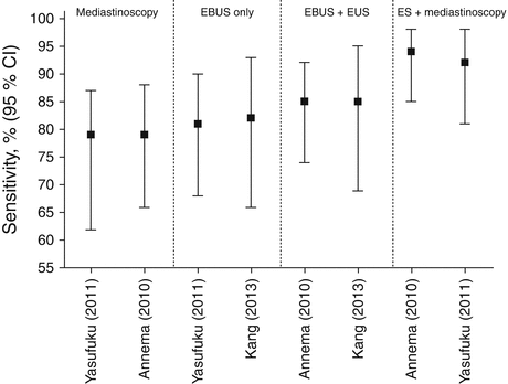
The predominant role of mediastinoscopy has been challenged by linear endobronchial and esophageal ultrasonography. Several meta-analyses on EUS-FNA alone, EBUS-TBNA alone, and combined EUS + EBUS reported a pooled sensitivity of 83–94 % for mediastinal staging of lung cancer [25–29]. Three controlled trials have been published so far. The two staging strategies proposed in the 2007 ESTS guidelines (mediastinoscopy alone versus alternatively combined linear endosonography followed by surgical staging whenever endosonography is negative) have been compared in a randomized controlled trial [30, 31] (Fig. 2.1). This trial showed that a staging strategy starting with combined (first esophageal approach using a dedicated EUS-scope, followed by the airway approach using the EBUS-scope) linear endosonography detected significantly (P = 0.02) more mediastinal nodal N2/3 disease compared to cervical mediastinoscopy alone, resulting in a significantly (P = 0.02) higher sensitivity of 0.94 (95 % CI 0.85–0.98) compared to 0.79 (95 % CI 0.66–0.88), respectively [31]. More recently, a meta-analysis on combined linear endosonography reported a negative likelihood ratio of 0.15, implying that the probability of having mediastinal nodal involvement for the individual patient with a negative combined linear endosonography result is 15 % [29]. This probability based on combined linear endosonography alone is not low enough to directly proceed to an anatomical surgical resection. In other words, a preoperative surgical mediastinal staging procedure is still recommended in the routine clinical practice after a negative (or incomplete) combined linear endosonography. A second controlled trial performed and compared both linear endosonography (airway approach only, using an EBUS-scope) and cervical mediastinoscopy in 153 patients with resectable stage I-III lung cancer [32]. There was no significant difference in sensitivity or negative predictive value, but the prevalence of mediastinal nodal disease in this study was lower than in most other studies and it should not be forgotten that in 29 % of mediastinal nodal stations an inadequate sample was obtained by EBUS-TBNA [32]. The sensitivity and negative predictive value for mediastinoscopy compared to EBUS-TBNA were 0.79 (95 % CI 0.62–0.87) versus 0.81 (95 % CI 0.68–0.90), and 0.90 (95 % CI 0.83–0.95) versus 0.91 (95 % CI 0.84–0.95), respectively [32]. A combined EBUS-TBNA and mediastinoscopy resulted in a sensitivity of 0.92 (95 % CI 0.81–0.98) and negative predictive value of 0.96 (95 % CI 0.90–0.99), or negative likelihood ratio of 0.04 [32]. Finally, a recent controlled trial did compare two strategies of combined (both the esophageal and airway route were performed using an EBUS-scope) linear endosonography by randomizing towards either an EUS centered approach (commencing with the esophageal route) or an EBUS centered approach (commencing with the airway route) [33]. There was no significant difference in diagnostic accuracy in both arms [33]. While the EBUS-centered approach didn’t result in statistical significant increase in accuracy or sensitivity (despite a stage shift in 7 % of patients for the second procedure), the EUS-centered approach resulted in a significant increase in diagnostic accuracy and sensitivity when an EBUS route was added (responsible for a stage shift in 48 % of patients). This study suggested that EBUS-TBNA is the better primary procedure in combined linear endosonography for mediastinal nodal staging of resectable stage I-III lung cancer.


Fig. 2.1
Across controlled trial comparison of sensitivity for mediastinal nodal staging by mediastinoscopy alone, EBUS-TBNA alone, combined linear endosonography, or combined linear endosonography plus mediastinoscopy in resectable stage I-III non-small cell lung cancer. ES combined linear endosonography. Yasufuku et al. [32], Annema et al. [31], Kang et al. [33]
The implementation of endosonography for baseline mediastinal nodal staging clearly reduces the need for an invasive surgical mediastinal nodal staging (mainly mediastinoscopy) by >50 % in patients with resectable stage I-III lung cancer [31, 34]. The lower complication rate associated with thoracic endosonography is an additional argument for its use as the first invasive mediastinal staging procedure. EBUS-TBNA and EUS-FNA are safe procedures with reported minor complications in <1 % of cases [25, 26, 35].


