(1)
Ospedale Civile di Vigevano, Vigevano, Italy
This group of arrhythmias includes several specific syndromes, the most important of which are:
1.
The long QT syndrome (LQTS);
2.
The short QT syndrome;
3.
The Brugada syndrome.
The Long QT Syndrome
The LQTS is a congenital disorder characterized on the surface electrocardiogram (ECG) by abnormal prolongation of the QT interval. The syndrome is caused by genetic mutations that alter the function of transmembrane sodium, potassium, and calcium ion channels, and it is associated with an increased risk of potentially fatal ventricular arrhythmias. The most characteristic is torsade de pointes (TdP)-type ventricular tachycardia (Fig. 6.62), which often causes episodes of syncope but can also lead to sudden death in children and adolescents.
Two main patterns of genetic transmission have been identified. The autosomal recessive form known as the Jervell-Lange Nielsen syndrome is characterized by QT interval prolongation plus deafness. The phenotypic features of the autosomal dominant form (Romano-Ward syndrome) are the same as those of the recessive form without deafness. Over the past decade, at least ten genes have been identified as causes of LQTS. On the basis of genetic backgrounds, six types of Romano-Ward syndrome and two types of Jervell-Lange Nielsen syndrome are now recognized. Two other rarer forms of LQTS have also been described although not without controversy: LQTS 7 (also known as the Andersen-Tawil syndrome), which is associated with skeletal abnormalities, and the LQTS 8 (or Timothy syndrome), which is associated with congenital heart disease, cognitive problems, and musculoskeletal diseases.
Thus far, ten electrocardiographic types of LQTS have been distinguished, mainly on the basis of the morphology of their T waves. The three most common types (LQTS 1, 2, and 3) are also distinguished by the settings in which the major arrhythmic episodes occur:
1.
In LQTS 1, the arrhythmia occurs after physical exertion;
2.
in LQTS2, it occurs with emotional stress or at rest;
3.
in LQTS3, during sleep or rest.
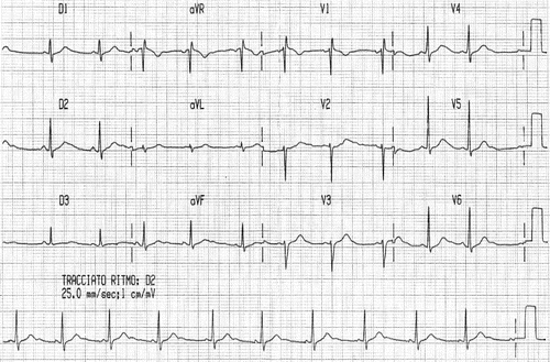
First-line screening for the LQTS obviously centers on the presence in the ECG of a long QT interval. Durations of <450 ms in males or <460 ms in females are considered normal (Fig. 9.1). It is important to recall that certain commonly used drugs are also capable of prolonging the QT interval. In patients with the LQTS, these drugs can provoke serious ventricular tachyarrhythmias and sometimes sudden death (Table 9.1).


Fig. 9.1
ECG in a patient with the LQTS showing prolongation of the QTc interval (560 ms) and T-U complex (kindly provided by Dr. Carla Giustetto)
Table 9.1
Drugs and the risk of torsade de pointes (TdP)
1. Drugs that increase the risk of TdP
Stay updated, free articles. Join our Telegram channel
Full access? Get Clinical Tree
 Get Clinical Tree app for offline access

|