and Muhammad A. Musani2
(1)
Department of Radiology, Stony Brook University Hospital, 100 Nicolls road, Stony Brook, NY, USA
(2)
Department of Medicine and Radiology, Stony Brook University Hospital, Stony Brook, NY, USA
Coronary artery CT evaluation is now widely performed on MDCT scanners with protocols optimized for high spatial resolution and reconstruction in various planes and fields of view [1]. Additionally, techniques may utilize contrast and thinner slice selection. This presents an opportunity to visualize and analyze other organs and structures besides the heart. Most commonly, the lungs, mediastinum, pulmonary vasculature, diaphragm, upper abdomen, and osseous as well as soft tissue structures of the thorax can be included in the field of view [2]. There has been a surge of reported extracardiac incidental findings that are discovered on coronary CT examinations and acknowledging them may be necessary as some are clinically significant and may require immediate action versus follow-up recommendations [3].
There is controversy surrounding the reporting of extracardiac incidental findings discovered on routine coronary CT protocols. Opponents argue that these incidental findings may lead to further investigations that would result in inappropriate resource utilization, increased healthcare costs, and increased patient anxiety [2]. Supporters argue that all data should be collected and if additional findings are indeterminate or significant, the physician has an ethical duty to act in the patient’s best interest [3].
Several technical factors will increase the prevalence of identifying incidental findings on routine interpretation of a coronary CT. The use of 64-slice MDCT coronary CTA, thinner slices, and advanced reconstruction algorithms provides superior spatial and temporal resolution and anatomic detail allowing for increased detection of incidental findings [4]. Another major contributing factor is the field of view. Volume analysis reveals that 35.5 % of the total chest volume is displayed when MDCT focuses on the heart as opposed to 70.3 % of the chest being visible when raw data is reconstructed with the maximum field of view [1].
Patient selection is another important consideration in regards to the probability of identifying significant incidental findings. Patients undergoing evaluation for coronary disease often times have risk factors and comorbidities that contribute to incidental findings, most commonly in the lungs. It has been reported that there is a significant correlation between any history of smoking and increasing age with the detection of extracardiac incidental findings which leads to imaging follow-up [5]. Given this correlation, it has been suggested that the entire chest be scanned on coronary MDCT for smokers older than 50 years, which may only add an additional 1 mSv of radiation. Interestingly, a statistically significant correlation has not been found between coronary calcium scores, age, and the detection of incidental findings [5]. Presenting symptoms may also increase the likelihood of incidental findings as a patient presenting with undifferentiated acute chest pain or shortness of breath may have extracardiac findings associated with their symptoms like aortic dissection or pulmonary embolism [6].
It has been reported that the incidence of incidental findings is 8 % in asymptomatic patients undergoing noncontrast chest CT for coronary artery calcium detection. Alternatively, the incidence may be as high as 58 % in patients with known or suspected coronary artery disease who receive a cardiac CTA, with up to 22 % requiring follow-up investigations [6]. The physician interpreting a cardiac CT must be familiar with potential incidental findings that can be encountered in order to identify them. Additionally, a determination must be made on whether a finding is benign, indeterminate, or significant. Then based on provided history and access to prior examinations, the physician must decide whether to report a certain finding and if follow-up investigations need to be recommended.
The most common extracardiac incidental finding reported during a cardiac CT evaluation is a pulmonary nodule. Recommendations for follow-up imaging of incidental solid (noncalcified) pulmonary nodules are usually based on established Fleischner Society guidelines [6]. Modified follow-up recommendation methodology has been proposed on cardiac CT, a 1-year follow-up examination for noncalcified nodules 2–5 mm in size, 6-month follow-up for nodules 6–9 mm in size, and immediate follow-up imaging for nodules larger than 9 mm [7].
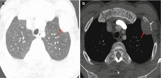
Clinically significant pulmonary incidental findings that require immediate follow-up include noncalcified nodules greater than 9 mm to 1.0 cm in size and lung masses, defined as size greater than 3.0 cm. Figure 9.1 shows a lung mass that is very concerning for cancer as it erodes the anterior left rib. There have been several cases of patients with shortness of breath undergoing coronary CTA who are found to have multiple pulmonary emboli, which often requires immediate attention [8]. Incidental discovery of pneumonia is critical to report. Figure 9.2 demonstrates a case of bilateral lower lobe pneumonia which follow-up to resolution imaging in 6–8 weeks post therapy should be recommended. Identification of a pneumothorax is extremely important, especially if the patient is undergoing cardiac CTA evaluation for chest pain, which will likely require changing of imaging protocols and initial management depending on size and complications [6].


Fig. 9.1
(a) Postcontrast CT axial image of the chest shows an extrapleural soft tissue mass in the anterior left upper chest measuring up to 4.0 cm in lung windows (red arrow) (b). Bone windows show the mass eroding the anterior left rib (white arrow). This mass is consistent with an aggressive lung cancer destroying the adjacent bone. The patient presented with chest pain. Follow-up recommendations included a complete contrast-enhanced chest, abdomen, and pelvis and possible CT-guided biopsy
< div class='tao-gold-member'>
Only gold members can continue reading. Log In or Register to continue
Stay updated, free articles. Join our Telegram channel

Full access? Get Clinical Tree


