16 Implications of Pediatric Renal, Endocrine, and Oncologic Disease
Renal
Hypertension
• Reduced compliance of the left ventricle (LV) secondary to LVH results in left ventricular diastolic dysfunction.
Key Points
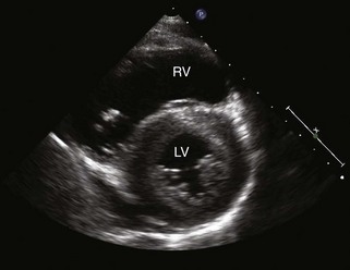
• Concentric hypertrophy occurs in the presence of pressure overload with a stiff ventricle (Fig. 16-1).
• Eccentric LVH is more likely secondary to increased volume overload (eg., in hemodialysis patients).
• Regression of LVH with normalization of left ventricular mass (LVM) can occur in patients after successful treatment with antihypertensive therapy.
Chronic Renal Failure
• African American race and female sex have been reported to be risk factors for cardiovascular disease in children and adolescents on dialysis.
Post-Kidney Transplantation
• Decreased systolic function, diastolic dysfunction, and increased LVM can persist even after transplantation.
The Echo Exam: Step-by-Step Approach
Step 1: Evaluate Cardiac Size
• M-mode and two-dimensional (2D) assessment of cardiac dimensions.
• Assess for left ventricular dilation and/or LVH using measurements indexed to body surface area (BSA).
• LVM equation is 0.8{1.04[(LVED + left ventricular posterior wall thickness + interventricular septal thickness)3 − LVED3]} + 0.6, where LVED is left ventricular end-diastolic dimension.
Step 2: Evaluate Systolic Cardiac Function
• Obtain end-diastolic and end-systolic measurements and calculate left ventricular fractional shortening (FS) using M-mode.
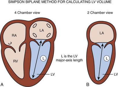
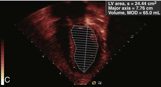
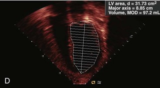
• Use the modified Simpson’s (disk summation) method for calculating ejection fraction (EF) to estimate left ventricular end-diastolic and end-systolic volumes.
Step 3: Evaluate Diastolic Cardiac Function
• Normal mitral inflow Doppler pattern.
• Isovolumic relaxation: brief interval between AV closure and the onset of ventricular filling (isovolumic relaxation time [IVRT]).
• In children with normal diastolic function, the E wave is larger than the A wave, reflecting that ventricular filling occurs primarily in early diastole.
• Increased HR results in shorter duration of diastole with an increased A wave and lower E/A ratio.
Stay updated, free articles. Join our Telegram channel

Full access? Get Clinical Tree


