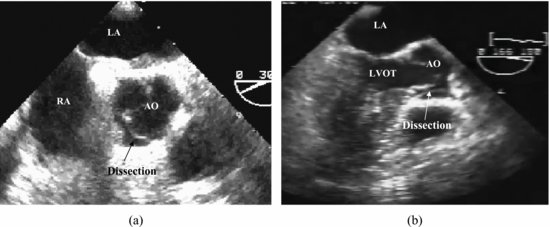
Figure 90.2 Transesophageal echocardiogram aortic short-axis view illustrates a dissection in the aortic root (a). Transesophageal echocardiogram long-axis view shows the dissection (b). AO, aorta; LA, left atrium; LVOT, left ventricular outflow tract; RA, right atrium.

Transthoracic echocardiography performed just after the accident and after 18 days showed neither aortic regurgitation nor dissection in the aortic root. PCI was successfully performed at the same day. Before the PCI, complete resolution of the dissection in the right coronary sinus was confirmed angiographically. Patient did well during a 8-month follow-up.
Discussion
Stay updated, free articles. Join our Telegram channel

Full access? Get Clinical Tree


