Fig. 12.1
(a, b) Axial transverse CT (a) and MPR reconstruction (curved) (b): partially thrombosed left common iliac artery aneurysm. With the growth in size (up to 32 mm), appeared an intermittent claudication related to the partial thrombosis of the left common iliac artery: the exclusion of the aneurysm with an iliac stent graft was planned, which involved first the occlusion of the ipsilateral hypogastric artery by coils, to avoid a later reinjection. (c) Aortography (25° RAO): short stenosis downstream of a proximally enlarged left common iliac artery. (d) Occlusion of the end of the hypogastric trunk by coils, via a distal ipsilateral femoral access. (e) Control aortography after deployment of a stent graft excluding the aneurysm and also treating the stenosis
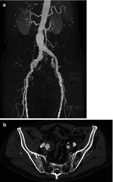
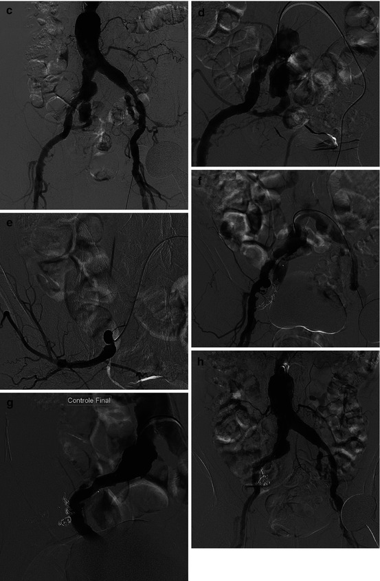
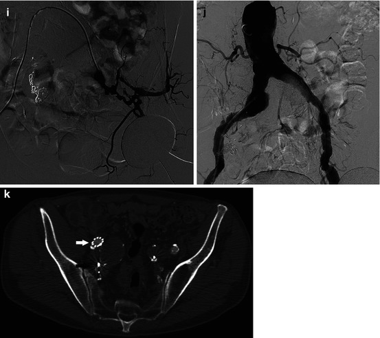
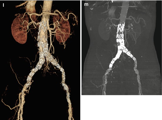
Fig. 12.2


Infrarenal aortic aneurysm extended to common iliac and hypogastric arteries. Embolization before bifurcated aortoiliac stent grafting (EVAR). (a) MIP reconstruction with calcified structures substraction showing the flowing lumen: infrarenal aortic aneurysm, respecting an infrarenal proximal neck, extended to the common iliac arteries; partially thrombosed internal iliac aneurysm. (b) Axial transverse CT scan through the origin of the two hypogastric arteries: partially thrombosed right hypogastric aneurysm, smaller left hypogastric aneurysm, partially flowing. (c) Aortography. (d) Right hypogastric aneurysm treatment: from a left femoral artery puncture, cross over to the right common iliac artery, and selective catheterization of the right hypogastric artery (Cobra catheter). (e–g) Hyperselective catheterization of the right gluteal artery (downstream of the aneurysm) (e), to be first excluded by coils + plug, to avoid any reinjection after the coverage of the common iliac aneurysm by a stent graft (f, g). (h) Control aortography via a right femoral access 3 weeks later: correct exclusion of the right hypogastric artery. (i) Selective catheterization of the left hypogastric artery from a right femoral access: after crossing over the aortoiliac bifurcation, injection before truncal plug exclusion, to avoid any reinjection after implantation of an aortoiliac stent graft. (j) Final aortographic control: exclusion of both hypogastric arteries, allowing the aorto-bi-iliac stent grafting (EVAR). (k–m) CT scan 1 month after the EVAR: axial scan (k), volume rendering, and MIP reconstruction (l, m): decrease in size of the internal iliac aneurysms, which are not enhanced. Right iliac limb of the stent graft: white arrow; coils at the end of the right hypogastric trunk and in the right gluteal artery (black arrow)
Stay updated, free articles. Join our Telegram channel
Full access? Get Clinical Tree
