Section 7 Hypertrophy patterns
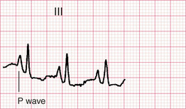
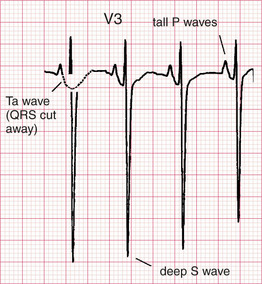
Right atrial abnormality (P-pulmonale)
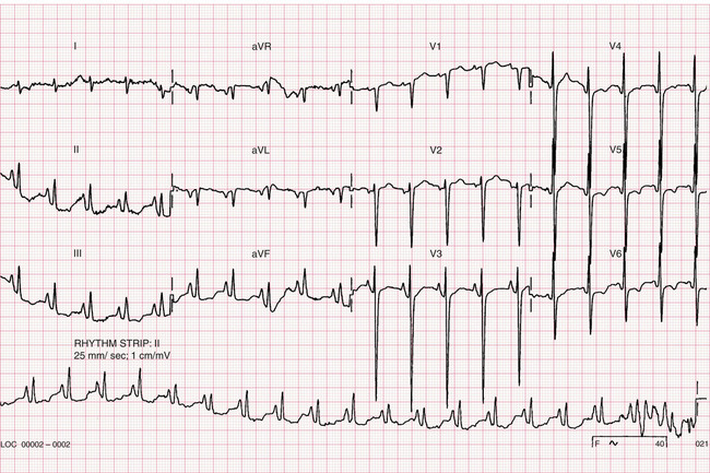
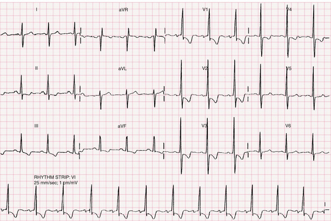
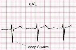
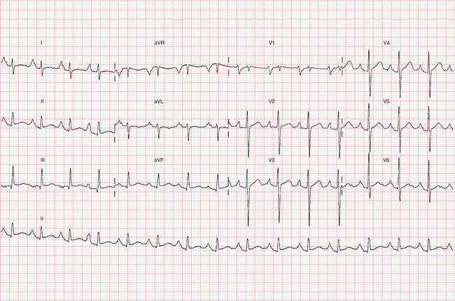
Features of this ECG
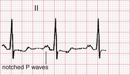
Left atrial abnormality (P-mitrale)
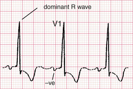
• Other features:
– prolonged duration (> 40 ms/1 small square) and increased amplitude (0.1 mV) of the terminal negative component to the P wave in V1.
Clinical Note
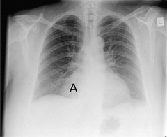
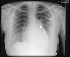
The combination of left atrial hypertrophy and right ventricular hypertrophy suggests mitral stenosis. This patient had mitral stenosis on the basis of rheumatic heart disease. His chest x-ray (Fig. 70.4) shows cardiomegaly, enlargement of the pulmonary outflow tract (A) and left atrial enlargement (B).
