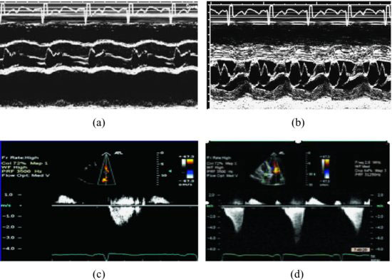
Figure 80.2 illustrates: The narrow LVOT causing the premature closure (arrow) of aortic leaflets. Systolic anterior motion (SAM) during systole by M-mode recording from aortic root and mitral inflow in a case with interventricular septum hypertrophy. Different shapes of continuous wave Doppler from LVOT are important indicators for diagnosis; normal is symmetric (Figure 80.2c) and asymmetric indicates obstruction of LVOT (Figure 80.2d). Three-dimensional echocardiography demonstrates the narrow LVOT and SAM (Figure 80.3).
Figure 80.2 M-mode recording from aortic root illustrates: the premature closure of aortic leaflets (a), the mitral anterior leaflet anterior motion during systole (systolic anterior motion) (b). Different shapes of continues wave Doppler from left ventricular outflow tract are important indicators for diagnosis; normal is symmetric (c) and asymmetric indicates the obstruction of left ventricular outflow tract (d).

Figure 80.3 Apical 3-chamber of the three-dimensional image illustrates the thickened septum caused left ventricular outflow tract stenosis and systolic anterior mitral leaflet anterior motion (arrow). AO, aorta; IVS, interventricular septum; LA, left atrium; LV, left ventricle; SAM, systolic anterior motion.
Stay updated, free articles. Join our Telegram channel

Full access? Get Clinical Tree


