5 Hypertensive Heart Failure
Left Ventricular Hypertrophy
How to Evaluate LV Mass
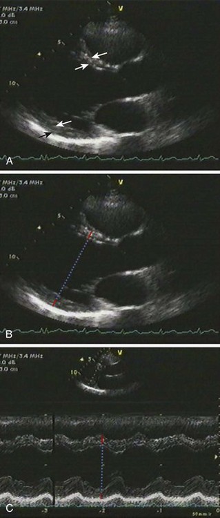
Linear Measurements
Key Points
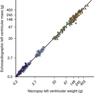
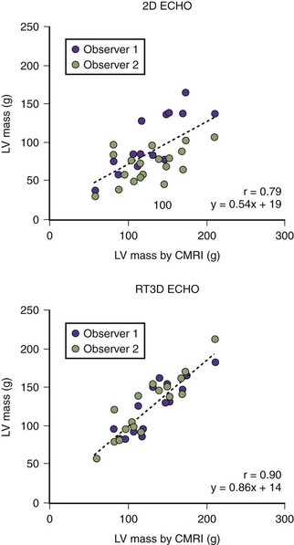
2D Measurement of LVM
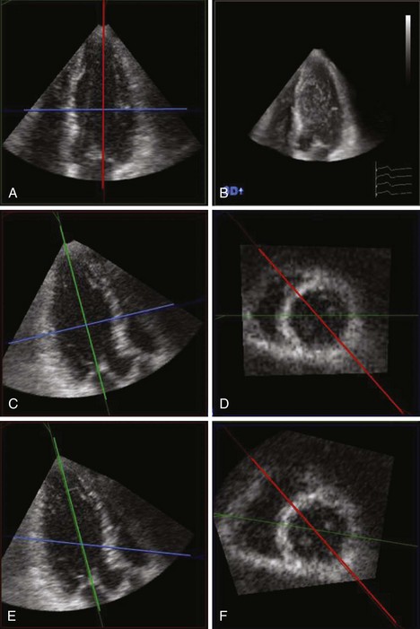
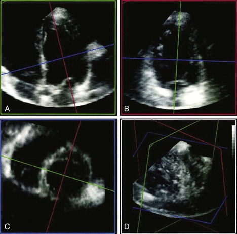
3D Measurement of LVM
Key Points
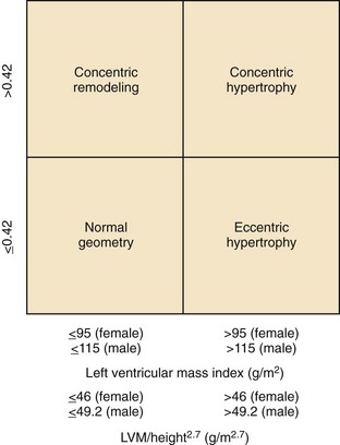
Definition of LVH and Abnormal LV Geometry
LV Hypertrophy
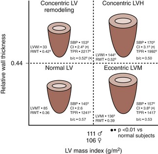
LV Geometry (Figure 5-8)
Key Points
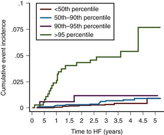
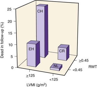
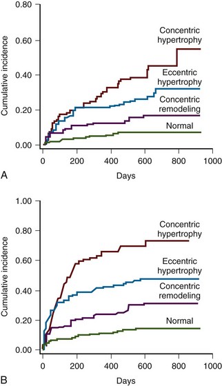
Figure 5-11 LVM and LVH raise the risk of incident heart failure events in the Multi-Ethnic Study of Atherosclerosis.
(Reprinted with permission from Bluemke DA, Kronmal RA, Lima JA, et al. The relationship of left ventricular mass and geometry to incident cardiovascular events: The MESA (Multi-Ethnic Study of Atherosclerosis) study. J Am Coll Cardiol. 2008;52:2148-2155.)
Clinical Vignettes
Case 1
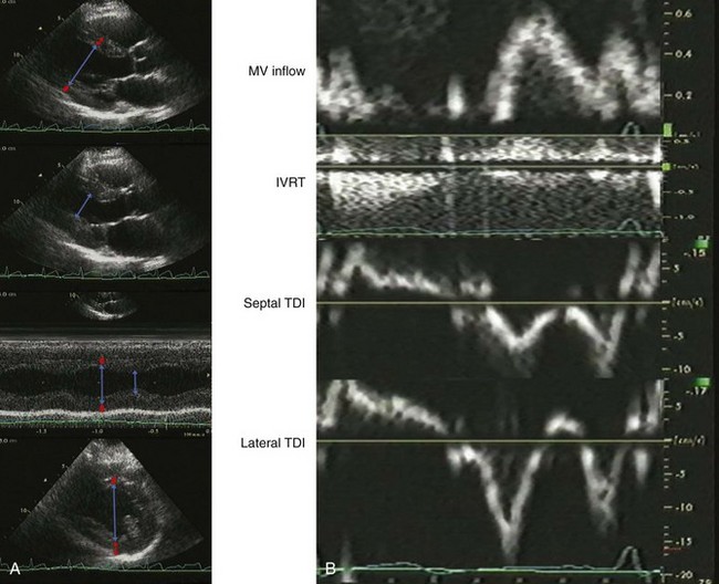
A 48-year-old diabetic, hypertensive man with unlimited exercise tolerance was referred to echocardiography to evaluate atypical chest pain. His blood pressure was 160/100 mm Hg on valsartan and hydrochlorothiazide. He was mildly overweight with a normal exam. The LV ejection fraction (LVEF) was normal with normal valvular function. On echocardiographic exam, LV geometry was normal (Figure 5-13A):
The mitral filling pattern was also normal (Figure 5-13B):
Case 2
A 60-year-old man with hypertension, myelofibrosis, and multiple falls underwent echocardiography during evaluation of presyncope. His blood pressure was 131/77 mm Hg on amlodipine and captopril. He was normal weight with a normal exam. His hemoglobin was 8.0 gm/dL. The LVEF was normal with normal valvular function (Figure 5-14A). On echocardiographic exam:
Stay updated, free articles. Join our Telegram channel

Full access? Get Clinical Tree


