Fig. 6.1
Arterial segmentation of the liver (selective injection of the CT via a humeral access, front and left oblique posterior view) (Courtesy Pr JL Lamarque)
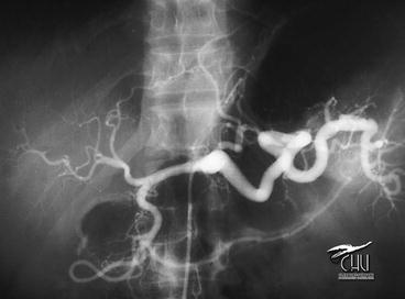
Fig. 6.2
Selective injection of the CT via a femoral access (Courtesy Pr JF Viallet)
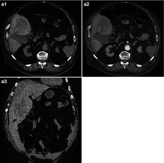
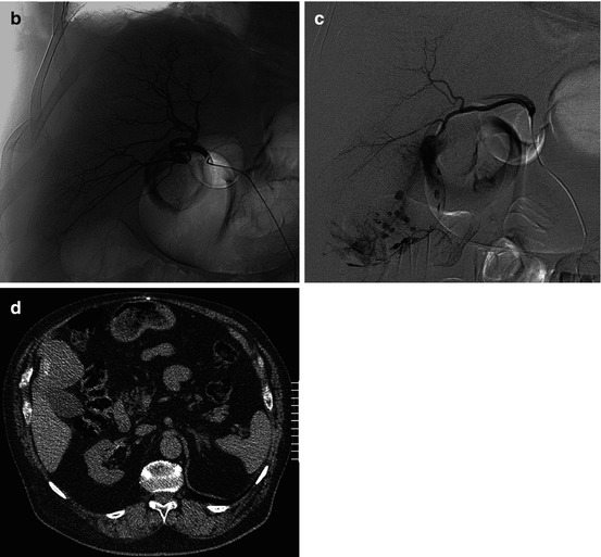
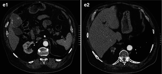
Fig. 6.3
68-year-old male, violent right hypochondrium pain: CT. (a) Subcapsular hematoma of the liver, heterogeneous segment V mass spontaneously hyperdense: these data and a clinical hypovolemic collapsus led to perform in emergency an angio. (b) Proper hepatic A selective injection: the liver is far from the abdominal wall; old costal fracture callus; heterogen hepatography. (c) Hyperselective segment VIII branch (arterial branch injection): massive extravasation, consequence of a hemorrhagic tumoral rupture; embolization using microparticles completed with Gelfoam. (d) Clinical aftereffects were simple; CT control 1 m later: residual hyperdensity inside a hypodense tumor. (e) Same CT: not any other tumor focus
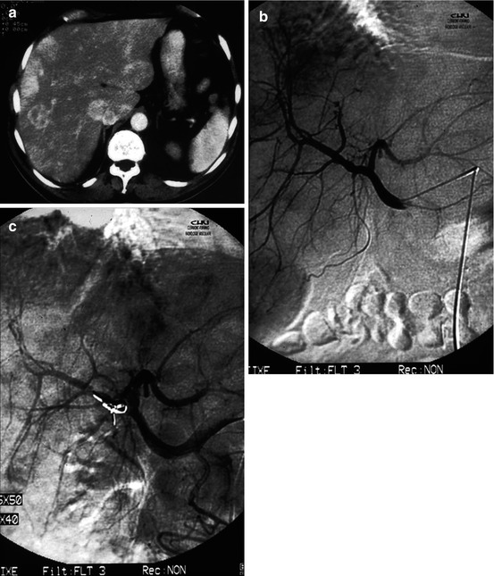
Fig. 6.4
Carcinoid secreting liver metastasis (primitive tumor unknown). Severe flushes led to decide an embolization to treat these symptoms. (a) Multiple hypervascular metastases. (b) Selective hepatic A injection. (c) CT scan after arterial tumoral-feeding microparticles embolization
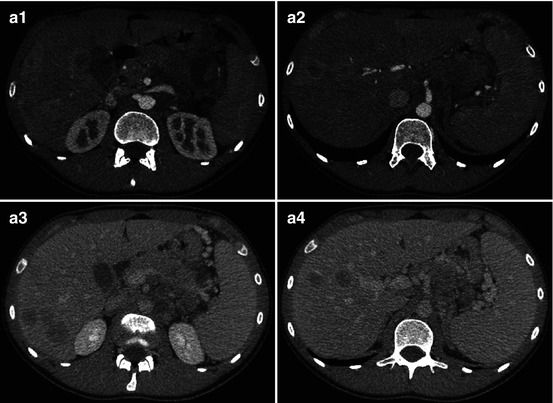
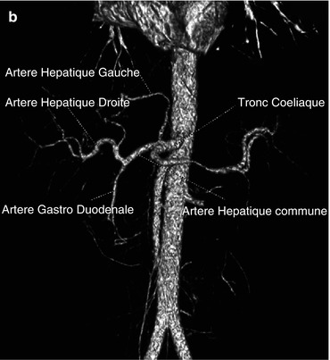
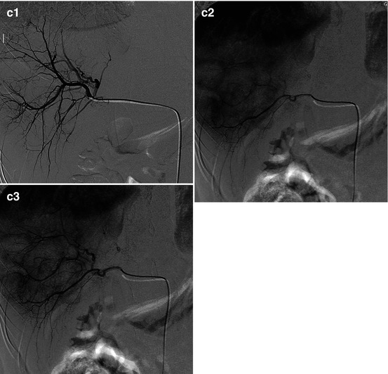
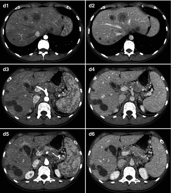
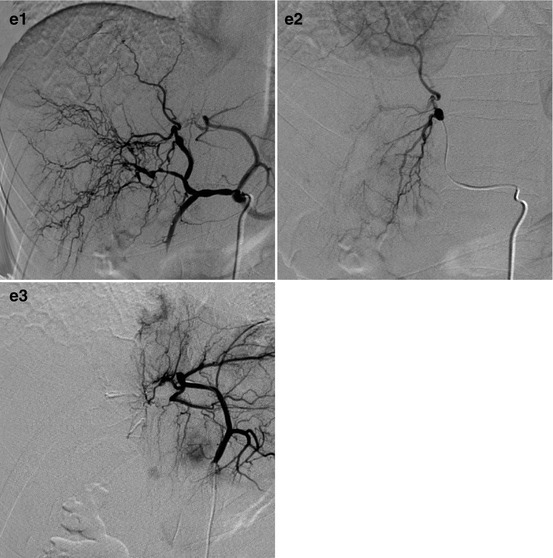
Fig. 6.5
21 years old, malignant pancreatic insulinoma surgically treated, multiple liver metastases. A complementary embolization has been decided to try to normalize the glycemia. (a) Multiple liver hypervascular metastases, among which some are heterogeneous. (b) CT angio: origin of the left hepatic A from the CT. (c) Selective catheterization of the right hepatic A, arterial and late phases: heterogeneous hepatography. Lateral and medial right liver sector particles embolization. (d) CT 5 weeks later: relative devascularization of the right masses; persistent segment IV hypervascularization and enhanced left liver metastasis. (e) Recurrence of hypoglycemias led to a re-embolization. Semi-selective injection of the CT: relative devascularization of the right liver, patent segment IV, and left hepatic A
Appendix: Liver Trauma: AAST Classification [9]
Gradea
Stay updated, free articles. Join our Telegram channel
Full access? Get Clinical Tree
 Get Clinical Tree app for offline access

|
|---|