Refractory epistaxes etiologies
3.1.2 Relevant Radiological Anatomy (Fig. 3.1)
A perfect knowledge of the branches of the carotid artery is an essential prerequisite. They must be located on conventional angiography after a selective catheterism of the external carotid artery.
The sphenopalatine artery, a branch of the internal maxillary artery, vascularizes the nasal septum and the lateral wall of the nasal fossae. Hence, it is the artery that is the most commonly involved in idiopathic epistaxes.
Some potential anastomoses between the internal and external carotid branches are at risk for an embolization and must be systematically looked for. These are:
Anterior ethmoidal arteries
Posterior ethmoidal arteries
Foramen lacerum arteries
Pterygoid arteries
Botallo’s foramen arteries
Foramen rotundum arteries
Angular arteries (a branch of the facial and ophthalmic artery)
Transclival branches of the carotid siphon
3.1.3 Epistaxes Treatment: Techniques and Indications
3.1.3.1 Initial Epistaxes Treatment Is Local
Manual compression using two fingers when treating a very anterior bleeding
Anterior gauze plugging using plugs (Merocel®)
Endoscopic electrocoagulation using bipolar forceps under local anesthetics
Anterior gauze plugging using inflatable balloons (e.g., Bivona®, Portex®)
Severe epistaxes are most commonly posterior epistaxis that induce oral cavity and nasal fossa bleeding. They should be treated by anterior and posterior nasal packing using a double-balloon probe. However, packing can be inefficient in a number of cases (25–52 % of cases according to series). In such cases, it is necessary to resort to surgery or to an endovascular treatment.
3.1.3.2 Surgical Treatment of Epistaxes
Surgery can be useful in case of uncontrolled bleeding. Several types of surgery are possible:
Ligation of the external carotid artery by cervicotomy: this technique is abandoned except in exceptional cases and makes any future embolization impossible.
Ligation of the internal maxillary artery: via a vestibular approach; it requires specialized skills and is not suitable for emergency treatment.
Endonasal ligation of the sphenopalatine artery: this technique consists in clipping the sphenopalatine artery at its emergence (sphenopalatine foramen) in the posterior part of the nasal fossa. It is effective in 75–85 % of cases (even more when performed by a highly experienced radiologist). Its limits are deterioration of the mucosa, which makes visualization of the artery difficult, as well as abundant bleeding linked to a hemostasis disorder. It is only used in case of unilateral and idiopathic epistaxes.
Ligation of the ethmoidal arteries: most often, it is performed when embolization fails and control angiography depicts show a revascularization of the nasal cavity via the anterior or posterior ethmoidal artery. Ligation of the anterior ethmoidal artery is more often performed through an internal canthal surgical approach rather than an endonasal approach. Ligation of the posterior ethmoidal artery is performed in the case of a hemorrhagic recurrence despite ligation of the anterior ethmoidal artery.
Generally, severe epistaxes must be treated by using anteroposterior packing. In the case of persistent or life-threatening bleeding, or in an unfavorable etiological context, they will be treated through surgical or endovascular radiologic approach.
3.1.3.3 Endovascular Treatment (Fig. 3.2)
Essential anterior epistaxes only require an embolization treatment if the local and/or surgical treatments fail.
Posterior epistaxes are a good indication of embolization if two posterior packings in 48 h are ineffective and if deglobulinization is important (hemoglobinemia under 8 g/dl).
Embolization technique: femoral approach using a valve introducer, then selective catheterism of the primitive carotid artery (if possible using a carrier catheter), internal and external homolateral to the bleeding (if necessary contralateral if the bleeding side is not identified during the ENT examination), and then microcatheterism (0.021 in.) of the target artery.
The arterial architecture and the bleeding area must be identified on frontal and lateral angiograms. Anastomoses with the arterial territories of the brain and ophthalmic arterial territories must be imperatively identified as well as anastomoses between the sphenopalatine and anterior ethmoidal arteries via turbinal and infraorbital arteries.
Catheterism of cervical arteries (subclavian artery and its branches) usually is not necessary in the case of idiopathic epistaxes. However, it is essential in other causes of ENT hemorrhages, especially postoperative in patients who have had a laryngectomy with blood on the tracheostomy tube.
Hemorrhages of the anterior ethmoidal artery require surgery and should not be treated with an endovascular approach because of the risks of ophthalmic artery microcatheterism.
Ipsilateral embolization of the sphenopalatine artery is sufficient in most cases. It can be associated with an embolization of the homolateral facial artery, which is often anastomosed with the sphenopalatine artery via the infraorbital artery. In the case of bilateral epistaxes, both sphenopalatine and both facial arteries can be embolized.
In some cases, the other branches of the external carotid artery can take over the terminal branch of the sphenopalatine and internal maxillary arteries via countercurrent anastomoses. Sometimes they only appear after occlusion of the main truncus and must then be catheterized and occluded.
3.1.3.4 Occlusion Material
Occlusion material can either be microparticles or microcoils or glue.
Nonresorbable microparticles with a caliber higher than 500 μm have an excellent effectiveness. However, they must be avoided in the case of anastomoses between the sphenopalatine and anterior ethmoidal territories, particularly if these two territories take part in the epistaxis. The use of microparticles requires a free-flow injection and fluoroscopic control to avoid any backflow and to locate anastomoses that would appear around the area. These microparticles can be a source of complications during embolization of the facial artery (skin necrosis, pain).
The use of microcoils can be a good alternative with pushable coils or controlled-release coils. Their positioning must be distal within the hemorrhagic area. The main drawback of this technique lies in the fact that it permanently fills the artery and, in the event of a recurrence, occludes one of the possible targets for a re-embolization.
The use of cyanoacrylate glues (Glubran 2® or Histoacryl®) is delicate and must be performed by specifically trained radiologists [5]. This technique has an immediate efficiency and is only used when the condition is life-threatening. Onyx embolization is easier and exposes to less off-target embolizations than cyanoacrylates. It requires a good knowledge of the toxicity and delivery of the product [6].
3.1.4 Epistaxes Embolization: Practical Factsheet
Environment:
Multidisciplinary team in angiography room: ENT surgeon, anesthetist, and interventional radiologist
Indications:
Posterior epistaxis refractory to a 48-h proper medical treatment
Severe epistaxis with immediate life-threatening condition
Assessment of the patient’s situation:
In all cases
Laboratory tests: PT, APTT, INR, CBC, platelets, and blood grouping
Controlled hemorrhage:
Etiologic investigation and technique assessment of the feasibility: angioscan of the supra-aortic trunks from the aortic arch to the circle of Willis and CT scan of the facial bones
Hemodynamic instability uncontrolled by resuscitation:
Endovascular (embolization) or surgical treatment must be discussed with ENT surgeon and anesthetist.
In the angiography suite:
Informed and reassured patient in supine position with a head rest, sedation, intravenous infusion, and monitoring
An interventional radiologist, an assistant, and a radiology technician
Anesthetist for sedation and general anesthetics if necessary, for monitoring and correction of hemodynamic parameters
On the angiography table:
6F valved introducer. Discuss 35-cm-long introducer according to the morphology of the iliofemoral arterial axis. 6F guide catheter (Envoy®, 100-cm type) on 0.035” hydrophilic guidewire (Terumo® type) and anti-backflow valve.
Microcatheter (minimum length 135 cm) suited for embolization agents (if necessary compatible with Onyx®) with straight or preformed tip (45° or 90°) with two radiopaque markers if controlled-release coils are used. 45° and 90° curved hydrophile microguides. Introducers, catheters, and microcatheters must be rinsed and infused using pressure infusion bags.
On the trolley in the angiography room:
Microcoils corresponding to the microcatheter
Calibrated microparticles (500–700-μm pre-filled Luer-Lock syringe)
Hemostatic and resorbable porcine gelatin (Gelita-Spon® type)
Cyanoacrylates (Histoacryl® or Glubran2® type)
Ethylene vinyl alcohol copolymer (Onyx®), rarely used in emergency
“Common” Procedure
Femoral arterial approach, rinsed and perfused valved introducer, and rinsed and perfused guide catheter:
Injection in the primitive carotid artery after ipsilateral selective catheterism, with an evaluation of the jugular venous return on late acquisition phases
Frontal and lateral selective acquisitions after injection of the internal carotid artery (study of the ophthalmic and anterior ethmoidal arteries)
Frontal and lateral selective acquisitions including nasal fossae after injection of the external carotid artery
Identification of hemorrhagic site and of dangerous anastomoses: foramen lacerum artery, clival artery, pterygoid artery, Botello’s foramen artery, foramen rotundum artery, and ophthalmic artery
Use of a microguide and a microcatheter: the latter is positioned at the origin of the external carotid artery. Hyperselective microcatheterism of the target (by default sphenopalatine artery).
Use of arterial tracing: catheterization of external carotid branches with 0.035 guide should be avoided to prevent spastic phenomena.
In the case of a dangerous anastomosis, place the microcatheter beyond that anastomosis, but always consider the risk of backflow during embolization. If necessary, perform a proximal (truncal) embolization of the dangerous anastomosis with microcoils before using microparticles.
In the case of arterial spasm and to enable free-flow embolization, use an in situ intra-arterial infusion of nitrates (1 mg RISORDAN®), in agreement with the anesthetist.
Systematic post-procedure control angiography is obtained to detect collaterals and takeover of the target. If the facial artery takes over the sphenopalatine artery, the facial artery must be embolized by very distal microspheres or microcoils.
Final control will search for a revascularization of the nasal fossa, notably of the ethmoidal arteries.
Leave the femoral introducer in place as long as hemorrhagic and neurologic control is not clinically confirmed by the ENT team in the angiography room.
3.1.5 Embolization Results
Technical success of embolization is important: 80–88 % [7].
Complications can occur in 8–13 % of cases [7–9]. The most recent studies show better results, probably due to progress in the techniques, in the training of the radiologists, in the material, and in embolization agents (microcoils, microparticles, etc.).
Complications of endovascular treatment of an epistaxis include hemorrhagic recurrences but also reported cases of facial neuralgia, septal perforation, sinusitis, and otitis media. Systemic complications can also occur: post-inhalation hypoxia, hypovolemia, angina, and/or myocardial infarction. Cullen’s meta-analysis on 539 patients has made it possible not only to make an inventory of the various complications of endovascular treatment but also to compare its rate of occurrence to that of surgical treatment [10]. Among the complications of arterial embolization, cerebrovascular accidents and occlusion of the central retinal artery [11] are the most serious. The radiologists’ experience, the patient’s arterial condition, and the embolization agents are likely to play a determining role in its occurrence. Few authors have made a critical analysis of the material and embolization agents used for this indication. Systematic use of microcatheterism contributes to its reduction [12].
In terms of costs of the procedure, studies data are controversial, possibly because of the uneven price of the various embolization agents that can be used. It seems the technical cost of embolization is higher than that of endoscopic ligations of the sphenopalatine artery [13]. On the other hand, the length of hospitalization is shorter with embolization [14].
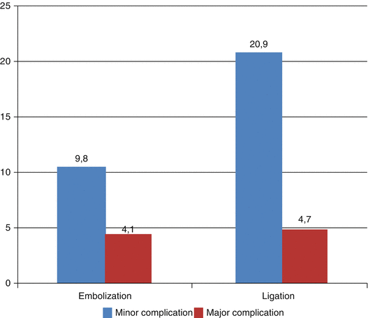
Comparison of minor and major complications of internal maxillary artery ligation versus embolization for refractory posterior epistaxis (Cullen [10])
3.1.6 Recurrence and Complications
Early rebleeding should be treated by embolization, except if ethmoidal arteries take over. Angiography will study collaterals (anterior ethmoidal, contralateral external carotid). Occlusion of both facial arteries and both sphenopalatine arteries is possible.
Stay updated, free articles. Join our Telegram channel

Full access? Get Clinical Tree


