Fig. 45.1
Left tracing, normal; right tracing typical of constriction. Normally there is a concordant fall in LV and RV pressures in inspiration. In constriction, there is discordance of LV and RV pressure changes in inspiration: the LV pressure falls, and the RV pressure rises
In cardiac tamponade, the physiological behavior is emphasized: right ventricular filling is maintained at the expense of restricted left ventricular filling. In inspiration, the increased volume of blood accommodated by the pulmonary vascular bed, coupled with reduced left ventricular filling, results in a greater reduction in systemic output [1].
45.2 Clinical Scenarios
45.2.1 Acute Pericarditis and Pericardial Effusion
There is a wide spectrum of causes of pericarditis associated with pericardial effusion or constrictive disease: infections (e.g., viral, bacterial, protozoal), immune-inflammatory disorders (systemic lupus erythematosus, rheumatoid arthritis, drugs), postradiation therapy, and neoplasia (primary and secondary) [1].
Clinical presentation is strongly influenced by the acute setting of the disease.
Physical examination: Regarding pericarditis, a pericardial friction rub is a typical finding, best heard when the patient is bent forward and accentuated at the end of expiration.
In tamponade, the combination of the classic findings known as Beck’s triad (hypotension, jugular venous distention, and muffled heart sounds) occurs in only 10–40 % of patients. Tachycardia, tachypnea, and hepatomegaly are common. Pulsus paradoxus (explained below) is relatively nonspecific and insensitive.
Pulsus paradoxus (or Kussmaul’s pulse): It is defined as a fall in arterial systolic pressure of >10 mmHg during normal inspiration.
Oximetry tracing often reveals the finding in the absence of direct arterial pressure line.
Diastolic pressure is not supposed to fall, thus reducing the difference between systolic and diastolic pressure with a consequent weakening of the pulse.
Beware that Kussmaul’s pulse is not specific for pericardial disease. It can occur in any condition with exaggerated inspiratory effort (pulmonary disease, pulmonary embolism, pleural effusion, congestive heart disease) [1]. It may also be obscured if the following conditions are also present: aortic insufficiency, atrial septal defect, and mechanical ventilation with positive end-expiratory pressure (PEEP). These are all cases that normalize the degree of left ventricular filling by favoring left ventricle volume load [2].
In the presence of tamponade, the right atrial and right ventricular pressure tracings reveal a blunted or an absent y descendant; hence there is no dip-and-plateau waveform (“square root sign”) and no Kussmaul’s sign.1
Jugular venous pressure: The x wave in the venous pressure trace is produced by atrial relaxation but predominantly by the systolic descent of the atrioventricular plane from ventricular contraction.
In cardiac tamponade, the x is steepened. The y is the result of early ventricular filling and lowering of the atrial venous pressure. It is prominent in conditions such as constriction and is attenuated in tamponade. In case of effusion–constriction, the x and y waves are usually similar producing an M or W pattern in right atrial tracing (Fig. 45.2). Also, this pattern is not specific for constrictive pericarditis but is also seen in heart failure, from restrictive cardiomyopathy and right ventricular infarction [2]. In constrictive disease, because of chronic elevation of right atrial pressure, hepatic congestion and dysfunction with ascites and peripheral edema are frequently encountered.
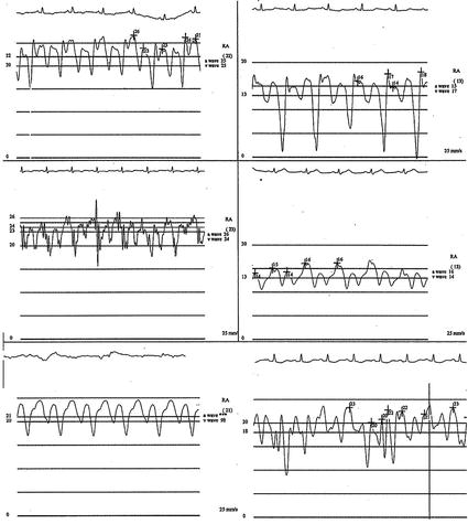
Fig. 45.2
Right atrial pressure in proven constriction cases. The mean pressures are all elevated. Although the x and y descent are all exaggerated, it can be seen how variable the depth of the x and y descent may be. In most cases, the magnitude of the x and y descents are similar, producing an M or W pattern. In the right upper tracing, the y descents are greatly dominant because of unusually vigorous recoil of the constrictive pericardium. In the right lower image, the inspiratory increase in x and y descents is depicted over two respiratory cycles. In later inspiration, the mean pressure rises as the right-sided heart compliance is exceeded by increased venous return and the pressure increases – Kussmaul’s sign. It is better appreciated by a mean pressure tracing
ECG findings: Typically, the electrocardiographic changes evolve through four stages characterized by diffuse ST-segment elevation and PR depression (seen in >80 % of patients), to normalization of the ST and PR segments, to widening of the T-wave. In cases where there is a moderate to severe pericardial effusion, low voltages may be seen and electrical alternans, a cyclic variation in QRS amplitude, when there is excessive motion of the heart within the fluid-filled pericardial space (swinging heart) [3].
Imaging: Echocardiography is the main diagnostic tool used for the diagnosis and characterization of the pericardial effusion, but it adds little where there is constrictive disease.
MRI is an optimal tool to study the morphologic characteristics of the pericardium and also allows the identification and characterization of other pathology [1].
45.2.2 Constrictive Disease
Constrictive physiology may develop after pericarditis. It is found in approximately 0.2 % of cases after open heart surgery, presenting a mean of 2 years postoperatively, and it is notable for occurring with underlying abnormal hearts (due to residual valve disease and/or infarction). Radiation therapy-induced constriction almost always displays concurrent fibrotic restrictive cardiomyopathy and fares far less well with surgical pericardiectomy than do other causes of constriction. In constrictive disease, the pericardium presents thickened, sometimes with calcification, but it can be apparently normal.
45.2.2.1 Clinical Scenarios
From the clinical point of view, symptoms are often vague and their onset is insidious; they include malaise, fatigue, and decreased exercise tolerance. Classical signs of right heart failure are typical (peripheral edema, nausea, abdominal discomfort, ascites).
Physical examination: Jugular venous distention is frequent and Kussmaul’s sign can be present even though it is sensitive but nonspecific for constriction. Auscultation reveals muffled heart sounds and occasionally a characteristic pericardial knock (60–200 ms after the second heart sound), caused by sudden termination of ventricular inflow by the encasing pericardium.
ECG findings: The ECG does not show specific findings, but low voltage may be seen.
Echocardiographic findings: Inflow Doppler analysis usually demonstrates mitral E wave reduction during inspiration due to inability of the left ventricle to generate a proper diastolic pressure because of the thick pericardium. The tissue Doppler findings will usually be normal excluding a myocardial muscle disease.
Cardiac catheterization: Ideally in constrictive pericarditis, a catheter study should be performed using mild sedation in order to minimize interference with respiratory physiology.
Arterial and venous accesses and two pressure transducers are needed to simultaneously record right and left pressures.
Required measurements include:
1.


Right atrial pressure
< div class='tao-gold-member'>
Only gold members can continue reading. Log In or Register to continue
Stay updated, free articles. Join our Telegram channel
Full access? Get Clinical Tree
