Hemodynamic Monitoring in Critical Illness

Chapter 35 Hemodynamic Monitoring in Critical Illness


The cardiovascular system is subject to rapid and profound changes during critical illness. Tissue hypoperfusion and hypoxia constitute an important cause of organ dysfunction in critically ill patients. Such pathophysiologic changes often are encountered in the setting of shock states attributable to inadequate cardiac output. Therapeutic maneuvers aimed at raising cardiac output have the potential to increase oxygen delivery to the whole body, as can be described by the simplified oxygen flux equation:


image


where image 
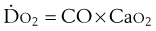
is whole-body oxygen delivery, CO is cardiac output, and CaO2 is arterial oxygen content. Increasingly, for the patient as a whole, flow is seen as a more important therapeutic target than pressure, although in certain organs, adequate perfusion pressure is known to be crucial (e.g., brain, heart, kidneys).


Monitoring of cardiovascular parameters allows clinicians to make informed decisions on how to optimize cardiac and circulatory function in order to maintain adequate tissue perfusion. Knowledge of the relevant anatomy and physiology is an essential prerequisite for correct interpretation of the data generated by any monitor.


Monitoring Devices


Central venous catheters, arterial catheters, and pulse oximeters are used routinely in the critical care setting. In addition, pulmonary arterial catheters, ultrasound-based cardiac output monitors, and devices that measure cardiac output by analysis of the morphology of the arterial pressure waveform also may be used. Despite advances in monitoring technology, significant complications are associated with their use, and to date, evidence for any consequent improvement in survival is lacking. This section addresses the indications, complications, and interpretation of data from the monitoring devices in common use.


Arterial Pressure Monitoring


Systolic, mean, and diastolic systemic arterial pressures are routinely measured. Mean arterial pressure (MAP) is a function of cardiac output (CO) and systemic vascular resistance (SVR).


image


Because vascular tone and thus SVR are independently controlled, it is not possible to use mean arterial pressure values alone to make assumptions about cardiac output. An adequate systemic blood pressure does not equate to adequate cardiac output or adequate flow in discrete tissue beds.


The systemic arterial pulse wave is generated in the left ventricle and is transmitted through the arterial tree at 6 to 10 meters/second. It comprises an incident pressure wave (from the contraction of the left ventricle) and a reflected pressure wave (from the periphery). As the pulse wave advances through the vascular tree, the systolic pressure is seen to increase as a result of an increase in the magnitude of the reflected wave. Therefore, measured systolic arterial pressure varies depending on the site of measurement. Mean arterial pressure may be a more useful marker than systolic blood pressure, because its value is less dependent on site of measurement, is least altered by damping, and is more relevant in determination of blood flow to vital tissue beds such as the brain and kidneys.


Noninvasive Arterial Blood Pressure Monitoring


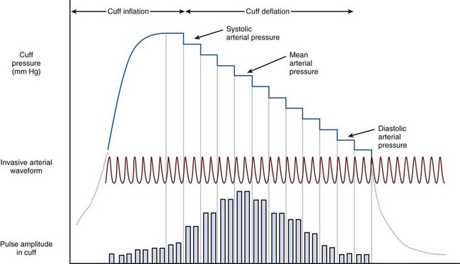
Noninvasive arterial blood pressure (NIBP) measurements in critical care most commonly are taken using an automated oscillometric device. This consists of a circumferential pneumatic cuff applied to the arm or leg. The cuff is inflated to a pressure above systolic arterial pressure, followed by controlled slow deflation of the cuff. As the cuff pressure falls below systolic arterial pressure, turbulent flow occurs in the artery beneath the cuff, causing oscillations in cuff pressure, which become maximal at MAP (Figure 35-1). Processing software allows determination of systolic, mean, and diastolic pressures in accordance with the amplitude of these oscillations.


This technique allows frequent measurement of blood pressure and can be used when continuous monitoring of arterial pressure is not required. It also provides useful confirmation of the reliability of invasive arterial measurement. Measurements in the normotensive range are considered to be accurate but tend to underestimate hypertensive values and overestimate hypotensive values. Measured values are less accurate in the presence of arrhythmia and with incorrect cuff sizing. Cuff width should be 20% greater than arm diameter, with use of a narrow cuff associated with a tendency toward erroneously high values.


Relative contraindications to cuff use include severe peripheral vascular disease, arteriovenous fistulas, local absence of lymph nodes consequent to resection, and local skin or muscle damage.


Complications are rare, but obtaining repeated measurements over short periods may lead to local skin ulceration or bruising. Injury to the ulnar nerve also has been reported.


Invasive Arterial Pressure Monitoring


Under conditions of hemodynamic instability or during therapeutic manipulation of the cardiovascular system, intermittent monitoring of blood pressure provides insufficient clinical information. An indwelling arterial catheter allows direct and continuous measurement of arterial blood pressure, as well as graphical display of the arterial waveform. Arterial blood sampling from the catheter also can be performed, allowing information to be obtained about metabolic status and respiratory function.


The radial, brachial, axillary, femoral, and dorsalis pedis arteries can be used for cannulation, with the radial artery most commonly used. Use of a 20-gauge cannula is recommended to reduce the incidence of vessel occlusion; a cannula of this size also has the most favorable physical properties for accurate pressure measurement. Insertion is performed under conditions of strict asepsis using either direct cannulation or a modified Seldinger technique. Distal perfusion should be periodically assessed after cannulation to ensure that arterial occlusion has not occurred.


The arterial cannula is connected by a continuous column of saline to a pressure transducer which takes atmospheric pressure at the level of the right atrium as its zero reference point. The system is continuously flushed from a pressurized saline source at a rate of 2 to 3 mL/hour to prevent aggregation of thrombus and subsequent occlusion of the cannula. Modifications to the transducer system and maneuvers promoting formation of clots or air bubbles should be avoided, because these can cause loss of energy from the system and “damping” of the measured signal.


Complications of arterial cannulation are listed in Box 35-1.


Central Venous Catheterization


Central venous catheters allow measurement of central venous pressure (CVP) and provide vascular access for blood sampling and administration of vasoactive drugs or parenteral nutrition. Common sites for catheterization include the internal jugular vein, subclavian vein, and femoral vein, although the brachial and cephalic veins also may be used. The chosen site will depend on the patient’s anatomy and clinical condition and on the experience of the operator. Two dimensional (2D) ultrasound imaging is readily available in many units and can be used in conjunction with Doppler color flow studies to define the venous anatomy before the procedure. Ultrasound imaging also may be used during the procedure to provide real-time guidance. The U.K. National Institute for Health and Clinical Excellence recommends the use of 2D ultrasound guidance for central venous cannulation. Correct catheter placement is indicated by the characteristic central venous pressure waveform and the radiographic appearance of the line tip positioned at the level of the carina on a plain chest radiograph.


CVP is measured continuously via a pressure transducer, and the CVP waveform may be displayed graphically. The availability and falling cost of disposable transducers have rendered intermittent manometry measurements obsolete. In health, CVP correlates with right ventricular end-diastolic pressure and pulmonary artery occlusion pressure (PAOP). It can therefore be used as an indicator of preload. In critical illness, the normal relationship between right- and left-sided heart pressures may not be maintained. This discrepancy may be the result of many factors, including changes in ventricular compliance, pulmonary hypertension, or pulmonary embolism. Isolated measurements of CVP are poor markers of intravascular volume status; however, dynamic measurements may still be useful. A sustained rise in CVP after a fluid challenge implies that a further increase in preload may not provide an increase in cardiac output.


The complications of central venous cannulation are listed in Box 35-2. Despite advances in catheter technology such as antimicrobial coating, risk for microbial contamination is significant. The corresponding risk of catheter-related sepsis may be mitigated by minimizing the time during which the catheter remains in situ. The benefits of changing the catheter must be weighed against the risks associated with catheter reinsertion (Box 35-3).


Pulmonary Artery Catheterization


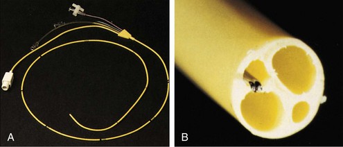
The pulmonary artery flotation catheter (PAFC) is a multilumen central venous catheter that is guided by flow to rest in the pulmonary artery (Figure 35-2). It allows measurement and derivation of a wide variety of hemodynamic variables, which are listed in Box 35-4. Indications for its use include the following:


Jun 12, 2016 | Posted by in RESPIRATORY | Comments Off on Hemodynamic Monitoring in Critical Illness

Full access? Get Clinical Tree

Get Clinical Tree app for offline access