Up to 44% isolated diastolic dysfunction, often mixed diastolic and systolic dysfunction
Most commonly secondary to ischemic heart disease (50%-75%) or primary valvular disease
Differential diagnosis for nonischemic dilated cardiomyopathy (DCM) is very broad including toxins, medications, autoimmune, viral and bacterial infection, nutritional, familial, endocrine, pregnancy, isolated ventricular noncompaction
Associated most commonly with chronic hypertension (HTN), left ventricular hypertrophy (LVH), and metabolic syndrome
Infiltrative etiologies include hemochromatosis, sarcoidosis, and amyloidosis
Can be seen acutely with ischemia as well as chronic coronary artery disease (CAD) after multiple myocardial infarctions (MI)
TABLE 9-1 Clinical presentations of heart failure | ||||||||||||||||||||||||||||||||||||||||||||||||||||||||||||||||||||||||||||||||||||||||||
|---|---|---|---|---|---|---|---|---|---|---|---|---|---|---|---|---|---|---|---|---|---|---|---|---|---|---|---|---|---|---|---|---|---|---|---|---|---|---|---|---|---|---|---|---|---|---|---|---|---|---|---|---|---|---|---|---|---|---|---|---|---|---|---|---|---|---|---|---|---|---|---|---|---|---|---|---|---|---|---|---|---|---|---|---|---|---|---|---|---|---|
| ||||||||||||||||||||||||||||||||||||||||||||||||||||||||||||||||||||||||||||||||||||||||||
Recent chest pains (CP) or symptoms of ischemia
Recent palpitations
History of congestive heart failure (CHF), CAD, HTN
Prior cardiac workup (i.e., echo, stress test, cardiac catheterizations)
Functional status/exercise capacity (e.g., flights of stairs or number of blocks before having to rest)
Changes in weight (e.g., clothes or rings not fitting recently), dependent edema, nocturia, paroxysmal nocturnal dyspnea (PND), orthopnea (most sensitive symptom of elevated pulmonary capillary wedge pressure (PCWP))1
Recent changes in medications or missed doses—be specific about timing
Recent changes in eating habits such as dining out, special events (e.g., holidays and weddings)
Elevated jugular venous pressure (JVP) or + hepatojugular reflex
Hepatomegaly or pulsatile liver
Dependent peripheral edema
Ascites
Parasternal heave
S3 or S4 at lower R sternal edge
Tachypnea, diaphoresis
Enlarged or displaced point of maximal impulse (PMI)
Early inspiratory rales (often not present in chronic failure) and hypoxemia
Decreased breath sounds with decreased tactile fremitus at lung bases
S3 (specific if present but not very sensitive) or summation gallop
Murmur/thrill suggestive of severe valvular regurgitation or septal defect
Always exclude ischemia/infarct as etiology of acute HF
Rule out with serial enzymes, as in acute myocardial infarction (AMI)
Low-grade troponin may be detectable and even expected with significantly elevated left ventricular end diastolic pressure (LVEDP), CKMB often negative, and enzymes do not follow the typical rise and fall seen in an acute coronary syndrome
Chronic renal failure is often associated with CHF, termed the “cardio-renal syndrome”
Acute renal failure often due to further worsening of cardiac output
Remember that decreased cardiac output (CO) is one cause of prerenal azotemia
Rising creatinine during treatment of acute HF is associated with worse in-hospital and long-term outcomes
Often “volume overload” presentations associated with creatinine improvement in the face of diuresis …? Improved CO versus lowering venous back pressure/congestion on kidneys
Most useful for excluding CHF as a contributor to clinical presentation
>150 pg/mL had a sensitivity and specificity of 85% and 83% and LR (+) of 5.3, LR (−) 0.18 for the diagnosis of heart failure in the Breathing Not Properly trial3
Elevated BNP in the setting of intact systolic function (nl EF) is highly suggestive of diastolic dysfunction5
BNP >400 pg/mL is virtually diagnostic of LV failure contributing to symptoms6
May be normal in cases of restrictive or constrictive physiology
Use caution in interpreting test in patients with chronic renal insufficiency (CRI), where brain natriuretic peptide (BNP) elevation may be exaggerated
Pro-NT BNP had a higher cutoff but similar operating characteristics7:
Optimal cutoff for age:
<50 = 450 pg/mL
50 to 75 = 900 pg/mL
>75 = 1800 pg/mL
above cutoffs yield a sensitivity of 90%, specificity of 84%
Rule out cocaine or amphetamine use as an etiology of heart failure in suspected patients
Useful for rapidly detecting ST segment elevation MI (STEMI) as a cause of new onset HF but beware “strain” can be an effect of HF
Arrhythmias such as A fib or high-grade A-V block as a precipitant of HF
Q waves and left bundle branch block (LBBB) are good predictors of systolic dysfunction. QRS >220 milliseconds portends a poor prognosis
Nonspecific intraventricular conduction delay >160 milliseconds suggests cardiomyopathy
May demonstrate pulmonary edema, cardiomegaly, pleural effusions
Useful for excluding other etiologies of symptoms
If rapid change in heart size, consider pericardial effusion/tamponade
Universally the single most important test in the evaluation of new onset HF
Rapidly differentiates among many etiologies listed above
Can be difficult to identify restrictive or constrictive physiology
Markers of diastolic dysfunction controversial and difficult in A fib
ALWAYS evaluate for ischemia in new-onset CHF, consider angiography as below
Useful for assessing viability of myocardium and potential for response to revascularization
Dobutamine echo may help determine response to aortic valve replacement (AVR) in critical aortic stenosis
May be helpful in titration of inotropes and vasodilators
No benefit in large-scale randomized trials
Definitely if evidence of acute ischemia/infarct as cause of HF
Consider in newly diagnosed systolic dysfunction as the gold standard for evaluation of coronary disease and potentially treatable lesions
Infrequently used in acute setting, can be useful for determining a cardiac or pulmonary cause of dyspnea when the etiology is unclear
Used in severe HF in making objective assessments about transplantation
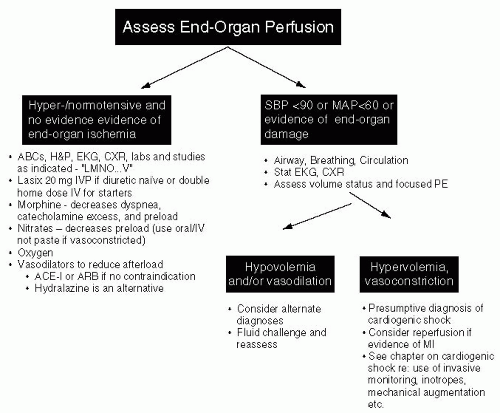
In a hemodynamically stable patient who is volume overloaded, your goal should be at least 1 to 2 liters/day
In patients with renal insufficiency, consider continuous furosemide infusion rather than bolus dosing8
Strict I/Os and/or daily weights are essential to monitor diuresis
Cardiac monitoring and frequent electrolyte checks (particularly magnesium and potassium) with aggressive replacement is essential to prevent arrhythmias
Encourage the patient to take an active role by asking the nursing staff their weight daily
Most patients respond to aggressive diuresis despite theoretical concerns of decreasing cardiac output
Diuretics alone rarely cause serious hypotension
Clinical deterioration with diuresis suggest preload-dependence such as severe pulmonary hypertension/RV failure, aortic stenosis, LV outflow tract obstruction, tamponade, constrictive pericarditis, or restrictive cardiomyopathy
If diuresis is inadequate consider adding metolazone (limited data),9 chlorothiazide, or nesiritide
Double the dose when switching from IV to oral furosemide to achieve equivalent diuretic effect
Consider oral bumetanide as an alternative to oral furosemide (better absorption in CHF but shorter elimination half-life)10
Consider continuous positive airway pressure (CPAP) as a bridge while diuresing and has recently been shown to have significant mortality benefit and decreases need for intubation in a large meta-analysis11
Strongly consider in patients with increased work of breathing or persistent hypoxia despite initial therapy
CPAP probably preferred to bilevel positive airway pressure (BIPAP) as there is some evidence of a trend toward increased AMI with BIPAP in comparison to CPAP11
Reasonable to start with 8 cm H2O and titrate to effect
Contraindications: Inability to protect airway, craniofacial deformities limiting mask fit, hemodynamic instability, high oxygen requirement that cannot be achieved with BIPAP, nausea/vomiting due to risk of aspiration, and significant arrhythmias
Optimize preload, minimize afterload via vasodilator and diuretic therapy
Prevent worsening of ejection fraction (EF) via ischemic events, tachyarrhythmia, excess afterload
Reduce risk of life-threatening arrhythmia
Multiple trials that show mortality benefit (see below). ACE-I12,13,14 or ARBs15 > nitrates and hydralazine16
ACE-Is are the preferred vasodilator and should be given a trial before hydralazine and nitrates
Enalapril, losartan, and candesartan best studied, but presumed to be class-effect
Start ACE-Is low and titrate while watching K and renal function closely
The lower the EF, the more benefit from ACE-I/ARB if tolerated
Allow for a 30% increase in creatinine with therapy before considering the patient intolerant
First vasodilator therapy to show survival benefit versus placebo16
Subsequently found to be inferior to ACE-I/ARBs,17 though best alternative for patients intolerant to ACE-I/ARB
Goal dose in VHeFT-I: hydralazine 300 mg/day + 160 mg isosorbide dinitrate/day16
May have an additive benefit to ACE-I/ARB, though only studied in self-identified “African descent” patients so far18


