Global Systolic Ventricular Function
Solomon Aronson1
Nhung T. Lam2
Solomon Aronson2
1OUTLINE AUTHOR
2ORIGINAL CHAPTER AUTHORS
▪ KEY POINTS
Assessment of global ventricular function is often the primary indication for perioperative echocardiography.
Transesophageal echocardiography (TEE) is well suited to providing accurate evaluation and monitoring of ventricular filling and systolic function.
Assessment of global ventricular function during hemodynamic instability is a class 1A indication for intraoperative TEE.
I. MEASUREMENT METHODS OF GLOBAL VENTRICULAR FUNCTION
A. Wall motion index (WMI)
Scaled scores are assigned to each segmental wall motion with the average providing a semiquantitative global assessment.
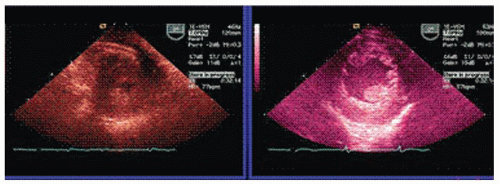
It has good agreement with estimated ejection fraction and other measures of ejection factor (Fig. 8-1).
Obtained from the transgastric short-axis view
The proportion of left ventricular (LV) chamber diastolic area in the mid-papillary short-axis view that is reduced during systole.
Fraction area change (FAC)% = [end diastolic area – end systolic area] × 100/end diastolic area
Often estimated visually
Chamber circumference can be traced in systole and diastole to provide more accurate calculation.
May miss wall motion abnormalities outside the one measured plane
Limited accuracy for assessment of overall ventricular function
Oblique planes may reduce accuracy.
B. Ejection fraction
Measure volume change (stroke volume) of the whole ventricle rather than area change of a single plane
More accurate measure of global ventricular function
EF% = [end diastolic volume – end systolic volume] × 100/end diastolic volume
Normal ejection fraction is 55% to 75%
C. Geometric methods (that fit a stereotypical ellipsoidal shape)
Single-plane ellipsoid method
Cylinder hemiellipsoid method
Area-length method
Estimate volume from diameter and length measurements in one or two planes.
Geometric assumptions limit accuracy of EF, when RWMA or unusual ventricular shapes are present
If measurement does not include the true apex (i.e., foreshortened view), then volumes and EF will also be unreliable.
Considered the best method for deriving ventricular volumes and ejection fraction
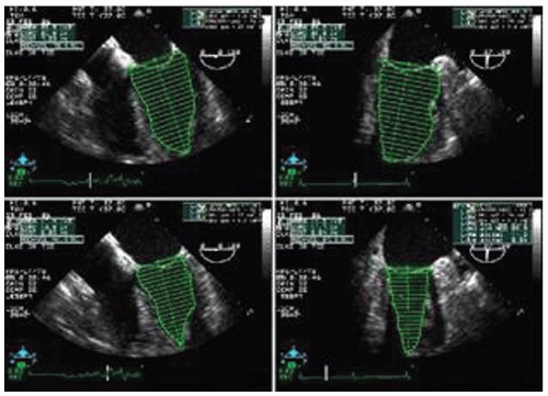
The endocardial border is traced in two orthogonal planes (e.g., midesophageal four-chamber and two-chamber views).
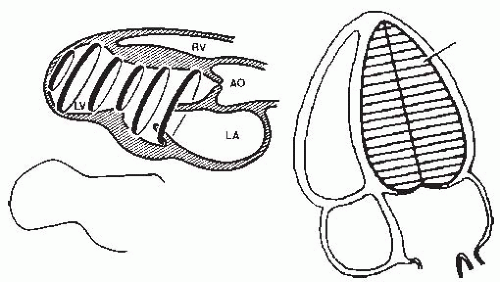
Computer software models the ventricle as a series of 20 or more stacked elliptical disks.
The volume of each disk is then calculated from the thickness of the disk, the diameters of each ellipsoid disk, and all of summed volumes to yield the total volume of the ventricle.
Cylindrical disks or rotating ellipsoid models can be generated from a single tomographic view, but with reduced accuracy, of one image plane (long axis).
Validated (angiography)
LV divided equal diameters
Cylindrical slices summed
E. The biplane disk summation method
Allows for variably shaped ventricles
Can account for significant regional wall motion abnormalities, but can still be limited by image quality or foreshortened views
Two views should not be combined if the chamber lengths differ by greater than 20% to reduce foreshortening errors.
TEE versus angiographic data is comparable (Table 8-1)1
Stay updated, free articles. Join our Telegram channel

Full access? Get Clinical Tree


